HitHunter cAMP 检测小分子试剂盒介绍

HitHunter cAMP 检测小分子试剂盒介绍

HitHunter cAMP 检测小分子试剂盒提供了一种稳健、高灵敏度且易于使用的检测方法,用于根据细胞中 3'-5'-环单磷酸腺苷 (cAMP) 的产生来监测 G 蛋白偶联受体 (GPCR) 的激活。该试剂盒非常适合筛选和分析小分子。对于测试生物制剂样品(例如血清或血浆中的粗制生物样品、纯化或未纯化的抗体[抗配体或抗受体]以及杂交瘤上清液),我们建议使用专为生物制剂设计的 HitHunter cAMP Assay for Biologics和大分子应用。其他来源的细胞系也与 HitHunter cAMP 检测兼容。本用户手册中的详细检测方案部分包含在 Gαs 受体检测中测试激动剂的程序、Gαi 激动剂、Gαs 和 Gαi 拮抗剂、抗配体抗体,以及测试 PAM 和 NAM 的建议。还包括适合高通量筛选的替代方案。

目录号 尺寸
90-0075SM2 200/800 dp(96 孔/384 孔)
90-0075SM10 1,000/4,000 dp(96 孔/384 孔)
90-0075SM25 2,500/10,000 dp(96 孔/384 孔)

Cell Meter 细胞活性检测试剂盒 *近红外荧光,适合微孔板检测*-AAT Bioquest荧光染料

上海金畔生物科技有限公司代理AAT Bioquest荧光染料全线产品,欢迎访问AAT Bioquest荧光染料官网了解更多信息。
Cell Meter 细胞活性检测试剂盒 *近红外荧光,适合微孔板检测*价格 2823
产品规格

200 Tests

产品货号

Cell Meter 细胞活性检测试剂盒 *近红外荧光,适合微孔板检测*

产品参数
Ex (nm) Em (nm)
分子量 溶剂
存储条件
产品概述

Cell Meter检测试剂盒是一类用于检测细胞功能的系列试剂盒,包括细胞活性、细胞毒性、细胞凋亡、细胞膜电位以及细胞周期等方面的指标。每种检测方案均能提供不同荧光颜色的检测方案。这些检测方案为从多角度研究细胞功能活动提供了一种十分有效的方法。Cell Meter细胞活性检测试剂盒 *近红外荧光,适合微孔板检测* 使用一种专属型染料,一旦进入活细胞中,其荧光强度增强。这种染料是一种疏水性复合物,可以轻易地进入完整的活细胞。通过细胞内酯酶水解弱荧光性底物,产生具有强烈荧光的亲水性产物,并且可以在保持在细胞质中。这种酯酶的活性与活细胞数量成比例关系,因此酯酶水解荧光底物形成的产物的荧光强度与活细胞数量相关。生长在培养板中的细胞可以被染料,且在不到两小时内就可以完成定量分析。本检测法比四唑盐或者基于Alarmar Blue的检测更加强大、稳定。它可以用于许多荧光实验平台的高通量分析中,例如微孔板分析,免疫组化和流式细胞术。它在许多研究中非常有用,包括细胞粘附性、趋化作用、药物抗性、细胞活性、细胞凋亡和细胞毒性研究。本试剂盒提供所有必需组分检测方案。它适合于增殖型和非增殖型细胞,也可用于悬浮和贴壁细胞。96微孔板中,每孔用试剂100ul,本试剂盒提供的试剂足以进行200次检测;384微孔板中,每孔加试剂25ul,本试剂盒提供的试剂足以进行800次检测。


适用仪器


荧光酶标仪  
Ex: 635 nm
Em: 670 nm
Cutoff: 665 nm
推荐孔板: 黑色透明底板
读取模式: 底读模式
实验方案

样品实验方案

简要概述

准备含测试化合物的细胞溶液
取出介质
加入CytoCalcein™NIR工作溶液(100 µL /孔/ 96孔板或25 µL /孔/ 384孔板)
在室温或37°C下孵育1小时
在Ex / Em = 635/670 nm(截止= 665 nm)时读取荧光强度(底部读取模式)


溶液配制

1.储备溶液配制

除非另有说明,否则所有未使用的储备溶液应分成一次性等分试样,并在制备后储存在-20°C。 避免反复冻融循环。
CytoCalcein™NIR储备溶液:
将20 µL DMSO(组分B)添加到CytoCalcein™NIR(组分A)的小瓶中,并充分混合以制成CytoCalcein™NIR储备液。 注意:20 µL CytoCalcein™NIR储备液足以装满一块板。 避光。 为了存放,请将管子密封。


2.工作溶液配制

将20 µL CytoCalcein™NIR储备溶液添加到瓶中的测定缓冲液(10 mL,组分C)中并充分混合以制成CytoCalcein™NIR工作溶液。 注意:此CytoCalcein™NIR工作溶液适用于一块细胞板,在室温下至少可稳定2小时。有关细胞样品制备的指南,请点击查看。


操作步骤

1.根据需要用测试化合物处理细胞。注意:添加化合物之前不必洗涤细胞。但是,如果测试的化合物对血清敏感,则可以在添加化合物之前将生长培养基和血清因子吸掉。加入100 µL /孔(96孔板)和25 µL /孔(384孔板)的1X Hank盐溶液和20 mM Hepes缓冲液(HHBS)或抽吸后选择的缓冲液。或者,细胞可以在无血清培养基中生长。

2.从细胞中除去培养基。注意:加载CytoCalcein™NIR工作溶液之前,必须除去培养基。

3.加入100 µL /孔(96孔板)或25 µL /孔(384孔板)的CytoCalcein™NIR工作溶液。

4.在室温或37°C下避光放置1小时。注意:适当的孵育时间取决于所用的单个细胞类型和细胞浓度。优化每个实验的孵育时间。对于非贴壁细胞,建议在孵育后以800 rpm的速度离心细胞板2分钟,然后制动。

5.使用荧光酶标仪(底部读取模式)在Ex / Em = 635/670 nm(截止= 665 nm)下监控荧光强度。


参考文献

Functional imaging of neuronal activity of auditory cortex by using Cal-520 in anesthetized and awake mice
Authors: Jingcheng Li, Jianxiong Zhang, Meng Wang, Junxia Pan, Xiaowei Chen, Xiang Liao
Journal: Biomedical Optics Express (2017): 2599–2610

NINJ2–A novel regulator of endothelial inflammation and activation
Authors: Jingjing Wang, Jingjing Fa, Pengyun Wang, Xinzhen Jia, Huixin Peng, Jing Chen, Yifan Wang, Chenhui Wang, Qiuyun Chen, Xin Tu
Journal: Cellular Signalling (2017)

Erythropoietin Stimulates Endothelial Progenitor Cells to Induce Endothelialization in an Aneurysm Neck After Coil Embolization by Modulating Vascular Endothelial Growth Factor
Authors: Peixi Liu, Yingjie Zhou, Qingzhu An, Yaying Song, Xi Chen, Guo-Yuan Yang, Wei Zhu
Journal: MEDICINE (2016): 1–8

Flexible Endoscopic Spray Application of Respiratory Epithelial Cells as Platform Technology to Apply Cells in Tubular Organs
Authors: Anja Lena Thiebes, Manuel Armin Reddemann, Johannes Palmer, Reinhold Kneer, Stefan Jockenhoevel, Christian Gabriel Cornelissen
Journal: Tissue Engineering Part C: Methods (2016): 322–331

Influence of hypothermia and subsequent rewarming upon leukocyte-endothelial interactions and expression of Junctional-Adhesion-Molecules A and B
Authors: Nicolai V Bogert, Isabella Werner, Angela Kornberger, Patrick Meybohm, Anton Moritz, Till Keller, Ulrich A Stock, Andres Beiras-Fernandez
Journal: Scientific reports (2016)

Inhibition of ABC transport proteins by oil sands process affected water
Authors: Hattan A Alharbi, David MV Saunders, Ahmed Al-Mousa, Jane Alcorn, Alberto S Pereira, Jonathan W Martin, John P Giesy, Steve B Wiseman
Journal: Aquatic Toxicology (2016): 81–88

Rapid generation of collagen-based microtissues to study cell–matrix interactions
Authors: Marie-Elena Brett, Alexandra L Crampton, David K Wood
Journal: Technology (2016): 1–8

Toxicokinetics and toxicodynamics of chlorpyrifos is altered in embryos of Japanese medaka exposed to oil sands process-affected water: evidence for inhibition of P-glycoprotein
Authors: Hattan A Alharbi, Jane Alcorn, Ahmed Al-Mousa, John P Giesy, Steve B Wiseman
Journal: Journal of Applied Toxicology (2016)

Spraying respiratory epithelial cells to coat tissue-engineered constructs
Authors: Anja Lena Thiebes, Stefanie Albers, Christian Klopsch, Stefan Jockenhoevel, Christian G Cornelissen
Journal: BioResearch open access (2015): 278–287

The efficient hemostatic effect of Antarctic krill chitosan is related to its hydration property
Authors: Shuai Wu, Zhuoyao Huang, Jianhui Yue, Di Liu, Ting Wang, Pierre Ezanno, Changshun Ruan, Xiaoli Zhao, William W Lu, Haobo Pan
Journal: Carbohydrate polymers (2015): 295–303


相关产品

产品名称 货号
Cell Meter 细胞活性检测试剂盒 红色荧光 Cat#22783
Cell Meter 细胞活性检测试剂盒 蓝色荧光 Cat#22785
Cell Meter 细胞活性检测试剂盒 蓝色荧光 405nm激发 Cat#22784

Cell Meter 磷脂酰丝氨酸凋亡检测试剂盒 蓝色荧光,适合微孔板检测-AAT Bioquest荧光染料

上海金畔生物科技有限公司代理AAT Bioquest荧光染料全线产品,欢迎访问AAT Bioquest荧光染料官网了解更多信息。
Cell Meter 磷脂酰丝氨酸凋亡检测试剂盒 蓝色荧光,适合微孔板检测 价格 2823
产品规格

100 Tests

产品货号

Cell Meter 磷脂酰丝氨酸凋亡检测试剂盒 蓝色荧光,适合微孔板检测

产品参数
Ex (nm) Em (nm)
分子量 溶剂
存储条件
产品概述

我们的Cell Meter™检测试剂盒是一套用于监测细胞活性的试剂盒。有多种参数可用于监测细胞活力。该特定试剂盒旨在通过测量磷脂酰丝氨酸(PS)的转运来监测细胞凋亡。在细胞凋亡中,PS转移到质膜的外部小叶。磷脂酰丝氨酸在细胞表面的出现是细胞凋亡的初始/中间阶段的通用指标,可以在观察形态变化之前进行检测。该特定试剂盒旨在通过测量磷脂酰丝氨酸(PS)的转运来监测细胞凋亡。该试剂盒使用我们专有的基于荧光小分子的Apopxin™PS探针,该探针以比膜联蛋白V(Kd <10 nM)高得多的亲和力特异性结合PS。与膜PS结合后具有蓝色荧光。它可以以酶标仪,显微镜和流式细胞仪的形式使用,而大多数其他商业凋亡检测试剂盒只能与显微镜或流式细胞仪平台一起使用。


适用仪器


荧光酶标仪  
Ex: 405 nm
Em: 450 nm
Cutoff: 420 nm
推荐孔板: 黑色透明底板
读取模式: 底读模式
实验方案

样品实验方案

简要概述

1.用测试化合物制备细胞(100 µL /孔/ 96孔板或25 µL /孔/ 384孔板)
2.加入等量的Apopxin™Violet 450工作溶液
3.在室温下孵育1小时
4.观察Ex / Em = 405/450 nm(截止= 420 nm)的荧光强度(底部读取模式)或使用装有紫罗兰色滤光片的荧光显微镜


溶液配制

工作溶液配制

将10μL100X Apopxin™紫罗兰色450(组分A)添加到1 mL的测定缓冲液(组分B)中,并充分混合以制成Apopxin™紫罗兰色450工作溶液。 


操作步骤

1.通过向PBS或所需的缓冲液中加入10X /孔(96孔板)或2.5 µL /孔(384孔板)的10X测试化合物储备溶液来处理细胞。对于空白孔(不含细胞的培养基),添加相同量的化合物缓冲液。
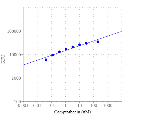
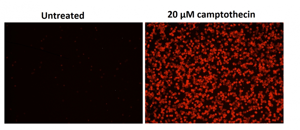
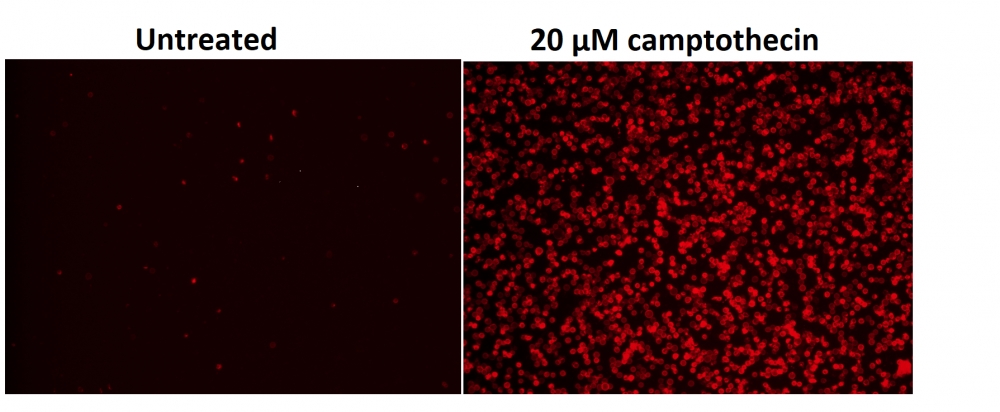
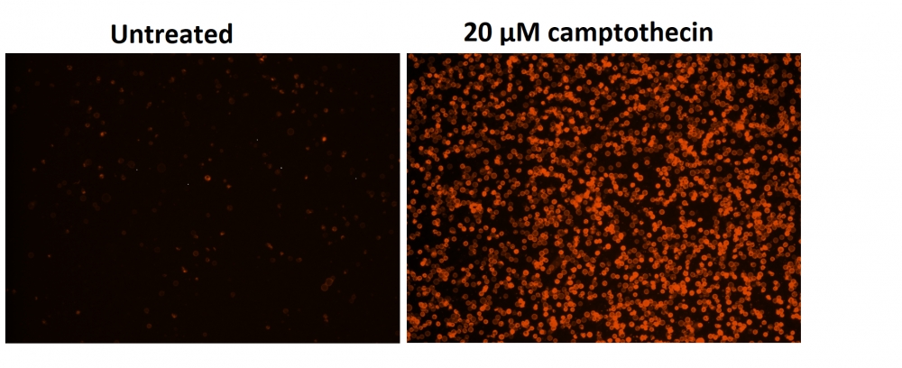
2.将细胞板在5%CO2、37°C​​的培养箱中孵育所需的时间(对于用星形孢菌素处理的Jurkat细胞需要4-6小时)以诱导凋亡。注意:由于在405 nm激发时,从380至490 nm的宽发射光谱,某些化合物(例如喜树碱)可能会产生假阳性反应。

3.在每个孔中添加100 µL /孔(96孔板)或25 µL /孔(384孔板)的Apopxin™Violet 450工作溶液。

4.在避光条件下,于室温下孵育细胞板至少1小时。

5.以800 rpm的速度离心细胞板(特别是非粘附细胞)2分钟(制动)。

6.使用荧光酶标仪(底部读取模式)在Ex / Em = 405/450 nm(截止= 420 nm)或使用带有紫色滤光片的荧光显微镜对图像池进行荧光强度监测。

Cell Meter 磷脂酰丝氨酸凋亡检测试剂盒 蓝色荧光,适合微孔板检测

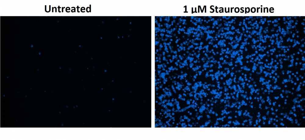
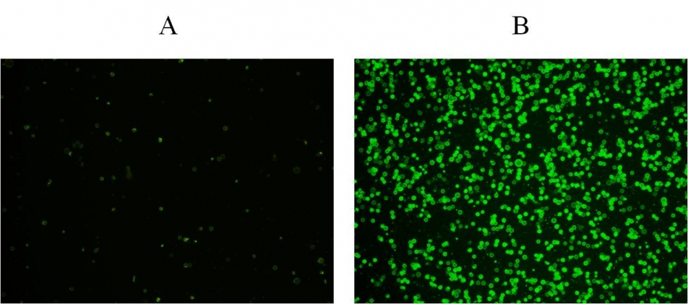
图1.用Apopxin™Violet 450偶联物染色的HeLa细胞的荧光图像。 Jurkat细胞在37℃下不使用(左)或用1μM星形孢菌素(右)处理4小时。 使用带有紫色滤光片组的显微镜(激发= 405nm)测量荧光强度。

参考文献

Physiological effects of the herbicide glyphosate on the cyanobacterium Microcystis aeruginosa
Authors: Wu, Liang and Qiu, Zhihao and Zhou, Ya and Du, Yuping and Liu, Chaonan and Ye, Jing and Hu, Xiaojun
Journal: Aquatic Toxicology (2016): 72–79

Tumor-selective mitochondrial network collapse induced by atmospheric gas plasma-activated medium.
Authors: Saito, Kosuke and Asai, Tomohiko and Fujiwara, Kyoko and Sahara, Junki and Koguchi, Haruhisa and Fukuda, Noboru and Suzuki-Karasaki, Miki and Soma, Masayoshi and Suzuki-Karasaki, Yoshihiro
Journal: Oncotarget (2016)

Inhibition of malignant phenotypes of human osteosarcoma cells by a gene silencer, a pyrrole–imidazole polyamide, which targets an E-box motif
Authors: Taniguchi, Masashi and Fujiwara, Kyoko and Nakai, Yuji and Ozaki, Toshinori and Koshikawa, Nobuko and Toshio, Kojima and Kataba, Motoaki and Oguni, Asako and Matsuda, Hiroyuki and Yoshida, Yukihiro and others
Journal: FEBS open bio (2014): 328–334

Detection of apoptosis based on the interaction between annexin V and phosphatidylserine
Authors: Liu T, Zhu W, Yang X, Chen L, Yang R, Hua Z, Li G.
Journal: Anal Chem (2009): 2410

Evaluation of cell surface expression of phosphatidylserine in ovarian carcinoma effusions using the annexin-V/7-AAD assay: clinical relevance and comparison with other apoptosis parameters
Authors: Dong HP, Holth A, Kleinberg L, Ruud MG, Elstr and MB, Trope CG, Davidson B, Risberg B.
Journal: Am J Clin Pathol (2009): 756

Mobilization of lysosomal calcium regulates the externalization of phosphatidylserine during apoptosis
Authors: Mirnikjoo B, Balasubramanian K, Schroit AJ.
Journal: J Biol Chem (2009): 6918

Peptidic targeting of phosphatidylserine for the MRI detection of apoptosis in atherosclerotic plaques
Authors: Burtea C, Laurent S, Lancelot E, Ballet S, Murariu O, Rousseaux O, Port M, V and er Elst L, Corot C, Muller RN.
Journal: Mol Pharm (2009): 1903

Suicidal membrane repair regulates phosphatidylserine externalization during apoptosis
Authors: Mirnikjoo B, Balasubramanian K, Schroit AJ.
Journal: J Biol Chem (2009): 22512

Trivalent methylated arsenical-induced phosphatidylserine exposure and apoptosis in platelets may lead to increased thrombus formation
Authors: Bae ON, Lim KM, Noh JY, Chung SM, Kim SH, Chung JH.
Journal: Toxicol Appl Pharmacol (2009): 144

Discovery of a phosphatidylserine-recognizing peptide and its utility in molecular imaging of tumour apoptosis
Authors: Thapa N, Kim S, So IS, Lee BH, Kwon IC, Choi K, Kim IS.
Journal: J Cell Mol Med (2008): 1649

Cell Meter 磷脂酰丝氨酸凋亡检测试剂盒 绿色荧光,适合微孔板检测-AAT Bioquest荧光染料

上海金畔生物科技有限公司代理AAT Bioquest荧光染料全线产品,欢迎访问AAT Bioquest荧光染料官网了解更多信息。
Cell Meter 磷脂酰丝氨酸凋亡检测试剂盒 绿色荧光,适合微孔板检测 价格 2823
产品规格

100 Tests

产品货号

Cell Meter 磷脂酰丝氨酸凋亡检测试剂盒 绿色荧光,适合微孔板检测

产品参数
Ex (nm) Em (nm)
分子量 溶剂
存储条件
产品概述

Cell Meter检测试剂盒是一类用于检测细胞功能的系列工具,包括细胞活性、细胞毒性、细胞凋亡、细胞膜电位以及细胞周期等方面的指标。每种检测方案均能提供不同荧光颜色的检测方案。这些检测方案为从多角度研究细胞功能活动提供了一种十分有效的方法。Cell Meter磷脂酰丝氨酸凋亡检测试剂盒 *绿色荧光,适合微孔板检测*是通过检测PS的翻转来测定细胞凋亡。在凋亡细胞中,PS从胞内移位到胞外表面,因此PS出现在细胞的外表面可以作为细胞凋亡的起始或者中间阶段的通用指示器,也可以通过其形态学改变来测定。该试剂盒使用我们独有的绿色荧光Apopxin PS传感器特异性的结合PS,其亲和性高于Annexin V(Kd<10nM)。该试剂盒使用的PS传感器结合膜上的PS后,发出绿色的荧光。因为与PS的高亲和性,该试剂盒比其它基于Annexin-V,仅能用荧光显微镜和流式检测凋亡的试剂盒更稳定,并且本试剂盒除上述两种仪器外还能使用荧光酶标仪。

点击查看细胞样品制备


适用仪器


荧光酶标仪  
Ex: 490 nm
Em: 525 nm
Cutoff: 515 nm
推荐孔板: 黑色透明底板
读取模式: 底读模式
实验方案

样品实验方案

简要概述

1.用测试化合物制备细胞(100 µL /孔/ 96孔板或25 µL /孔/ 384孔板)
2.加入等量的Apopxin™Green工作溶液
3.在室温下孵育1小时
4.在Ex / Em = 490/525 nm(截止= 515 nm)或带有FITC滤光片的荧光显微镜下监控荧光强度(底部读取模式)


溶液配制

工作溶液配制

将10 µL 100X Apopxin™Green(组分A)添加到1 mL的测定缓冲液(组分B)中,并充分混合以制成Apopxin™Green工作溶液。


操作步骤

1.通过向细胞制备缓冲液中加入10XL /孔(96孔板)的10X测试化合物溶液,以所需的测试化合物处理细胞,总体积为100μL/孔。对于384孔板,请在细胞制备缓冲液中使用5 µL /孔的5X测试化合物溶液,总体积为25 µL /孔。对于空白孔(不含细胞的培养基),添加相同量的化合物缓冲液。

2.将细胞板在5%CO2、37°C​​的培养箱中孵育所需的时间(对于喜树碱处理过的Jurkat细胞,需要4-6小时)以诱导凋亡。

3.在每个孔中添加100 µL /孔(96孔板)或25 µL /孔(384孔板)的Apopxin™Green工作溶液。

4.在避光条件下,于室温下孵育细胞板至少1小时。

5.以800 rpm的速度离心细胞板(特别是非粘附细胞)2分钟(制动)。

6.使用荧光酶标仪(Ext / Em = 490/525 nm(Cutoff = 515 nm))或使用带有FITC滤光片的荧光显微镜的图像池监控荧光强度。


图示

Cell Meter 磷脂酰丝氨酸凋亡检测试剂盒 绿色荧光,适合微孔板检测

图1.在Costar黑色壁/透明底部96孔板中,用Cell Meter™磷脂酰丝氨酸凋亡检测试剂盒染色的Jurkat细胞图像。 A:未经处理的对照细胞。 B:用20 µM喜树碱处理5小时的细胞。

参考文献

Physiological effects of the herbicide glyphosate on the cyanobacterium Microcystis aeruginosa
Authors: Wu, Liang and Qiu, Zhihao and Zhou, Ya and Du, Yuping and Liu, Chaonan and Ye, Jing and Hu, Xiaojun
Journal: Aquatic Toxicology (2016): 72–79

Tumor-selective mitochondrial network collapse induced by atmospheric gas plasma-activated medium.
Authors: Saito, Kosuke and Asai, Tomohiko and Fujiwara, Kyoko and Sahara, Junki and Koguchi, Haruhisa and Fukuda, Noboru and Suzuki-Karasaki, Miki and Soma, Masayoshi and Suzuki-Karasaki, Yoshihiro
Journal: Oncotarget (2016)

Inhibition of malignant phenotypes of human osteosarcoma cells by a gene silencer, a pyrrole–imidazole polyamide, which targets an E-box motif
Authors: Taniguchi, Masashi and Fujiwara, Kyoko and Nakai, Yuji and Ozaki, Toshinori and Koshikawa, Nobuko and Toshio, Kojima and Kataba, Motoaki and Oguni, Asako and Matsuda, Hiroyuki and Yoshida, Yukihiro and others
Journal: FEBS open bio (2014): 328–334

Detection of apoptosis based on the interaction between annexin V and phosphatidylserine
Authors: Liu T, Zhu W, Yang X, Chen L, Yang R, Hua Z, Li G.
Journal: Anal Chem (2009): 2410

Evaluation of cell surface expression of phosphatidylserine in ovarian carcinoma effusions using the annexin-V/7-AAD assay: clinical relevance and comparison with other apoptosis parameters
Authors: Dong HP, Holth A, Kleinberg L, Ruud MG, Elstr and MB, Trope CG, Davidson B, Risberg B.
Journal: Am J Clin Pathol (2009): 756

Mobilization of lysosomal calcium regulates the externalization of phosphatidylserine during apoptosis
Authors: Mirnikjoo B, Balasubramanian K, Schroit AJ.
Journal: J Biol Chem (2009): 6918

Peptidic targeting of phosphatidylserine for the MRI detection of apoptosis in atherosclerotic plaques
Authors: Burtea C, Laurent S, Lancelot E, Ballet S, Murariu O, Rousseaux O, Port M, V and er Elst L, Corot C, Muller RN.
Journal: Mol Pharm (2009): 1903

Suicidal membrane repair regulates phosphatidylserine externalization during apoptosis
Authors: Mirnikjoo B, Balasubramanian K, Schroit AJ.
Journal: J Biol Chem (2009): 22512

Trivalent methylated arsenical-induced phosphatidylserine exposure and apoptosis in platelets may lead to increased thrombus formation
Authors: Bae ON, Lim KM, Noh JY, Chung SM, Kim SH, Chung JH.
Journal: Toxicol Appl Pharmacol (2009): 144

Discovery of a phosphatidylserine-recognizing peptide and its utility in molecular imaging of tumour apoptosis
Authors: Thapa N, Kim S, So IS, Lee BH, Kwon IC, Choi K, Kim IS.
Journal: J Cell Mol Med (2008): 1649

Cell Meter 磷脂酰丝氨酸凋亡检测试剂盒 红色荧光,适合微孔板检测-AAT Bioquest荧光染料

上海金畔生物科技有限公司代理AAT Bioquest荧光染料全线产品,欢迎访问AAT Bioquest荧光染料官网了解更多信息。
Cell Meter 磷脂酰丝氨酸凋亡检测试剂盒 红色荧光,适合微孔板检测 价格 2823
产品规格

100 Tests

产品货号

Cell Meter 磷脂酰丝氨酸凋亡检测试剂盒 红色荧光,适合微孔板检测

产品参数
Ex (nm) Em (nm)
分子量 溶剂
存储条件
产品概述

Cell Meter检测试剂盒是一类用于检测细胞功能的系列工具,包括细胞活性、细胞毒性、细胞凋亡、细胞膜电位以及细胞周期等方面的指标。每种检测方案均能提供不同荧光颜色的检测方案。这些高效的检测方案为从多角度研究细胞功能活动提供了一种十分有效的方法。Cell Meter磷脂酰丝氨酸凋亡检测试剂盒 *红色荧光,适合微孔板检测*是通过检测PS的翻转来测定细胞凋亡。在凋亡细胞中,PS从胞内移位到胞外表面,因此PS出现在细胞的外表面可以作为细胞凋亡的起始或者中间阶段的通用指示器,也可以通过其形态学改变来测定。该试剂盒使用我们独有的红色荧光Apopxin PS传感器特异性的结合PS,其亲和性高于Annexin V(Kd<10nM)。该试剂盒使用的PS传感器结合膜上的PS后,发出红色的荧光。因为与PS的高亲和性,该试剂盒比其它基于Annexin-V,仅能用荧光显微镜和流式检测凋亡的试剂盒更稳定,并且本试剂盒除上述两种仪器外还能使用荧光酶标仪。

点击查看细胞样品配制


适用仪器


荧光酶标仪  
Ex: 590 nm
Em: 630 nm
Cutoff: 610 nm
推荐孔板: 黑色透明底板
读取模式: 底读模式
实验方案

样品实验方案

简要概述

1.用测试化合物制备细胞(100 µL /孔/ 96孔板或25 µL /孔/ 384孔板)
2.加入等量的Apopxin™Red工作溶液
3.在室温下孵育1小时
4.监视Ex / Em = 590/630 nm(截止= 610 nm)的荧光强度(底部读取模式)或使用带有Texas Red滤光片的荧光显微镜


溶液配制

工作溶液配制

将10 µL Apopxin™Red(组分A)加入1 mL的测定缓冲液(组分B)中,并充分混合以制成Apopxin™Red工作溶液。


操作步骤

1.通过向PBS或所需的缓冲液中加入10X /孔(96孔板)或2.5 µL /孔(384孔板)的10X测试化合物储备溶液来处理细胞。 对于空白孔(不含细胞的培养基),添加相同量的化合物缓冲液。

2.将细胞板在5%CO2、37°C的培养箱中孵育所需的时间(对于喜树碱处理过的Jurkat细胞,需要4-6小时)以诱导凋亡。

3.在每个孔中加入100 µL /孔(96孔板)或25 µL /孔(384孔板)的Apopxin™Red工作溶液。

4.在避光条件下,于室温下孵育细胞板至少1小时。

5.以800 rpm的速度离心细胞板(特别是非粘附细胞)2分钟(制动)。

6.使用荧光酶标仪(Ext / Em = 590/630 nm(截止= 610 nm))或使用带有TexasRed®滤光片的荧光显微镜对图像池进行荧光强度监测。


图示

Cell Meter 磷脂酰丝氨酸凋亡检测试剂盒 红色荧光,适合微孔板检测

图1.用Cell Meter™磷脂酰丝氨酸凋亡检测试剂盒染色的Costar黑壁/透明底部96孔板中Jurkat细胞的荧光图像。 不使用(左)或用20μM喜树碱(右)处理Jurkat细胞5小时。 使用具有Texas Red通道的荧光显微镜测量荧光强度。


参考文献

Physiological effects of the herbicide glyphosate on the cyanobacterium Microcystis aeruginosa
Authors: Wu, Liang and Qiu, Zhihao and Zhou, Ya and Du, Yuping and Liu, Chaonan and Ye, Jing and Hu, Xiaojun
Journal: Aquatic Toxicology (2016): 72–79

Tumor-selective mitochondrial network collapse induced by atmospheric gas plasma-activated medium.
Authors: Saito, Kosuke and Asai, Tomohiko and Fujiwara, Kyoko and Sahara, Junki and Koguchi, Haruhisa and Fukuda, Noboru and Suzuki-Karasaki, Miki and Soma, Masayoshi and Suzuki-Karasaki, Yoshihiro
Journal: Oncotarget (2016)

Inhibition of malignant phenotypes of human osteosarcoma cells by a gene silencer, a pyrrole–imidazole polyamide, which targets an E-box motif
Authors: Taniguchi, Masashi and Fujiwara, Kyoko and Nakai, Yuji and Ozaki, Toshinori and Koshikawa, Nobuko and Toshio, Kojima and Kataba, Motoaki and Oguni, Asako and Matsuda, Hiroyuki and Yoshida, Yukihiro and others
Journal: FEBS open bio (2014): 328–334

Detection of apoptosis based on the interaction between annexin V and phosphatidylserine
Authors: Liu T, Zhu W, Yang X, Chen L, Yang R, Hua Z, Li G.
Journal: Anal Chem (2009): 2410

Evaluation of cell surface expression of phosphatidylserine in ovarian carcinoma effusions using the annexin-V/7-AAD assay: clinical relevance and comparison with other apoptosis parameters
Authors: Dong HP, Holth A, Kleinberg L, Ruud MG, Elstr and MB, Trope CG, Davidson B, Risberg B.
Journal: Am J Clin Pathol (2009): 756

Mobilization of lysosomal calcium regulates the externalization of phosphatidylserine during apoptosis
Authors: Mirnikjoo B, Balasubramanian K, Schroit AJ.
Journal: J Biol Chem (2009): 6918

Peptidic targeting of phosphatidylserine for the MRI detection of apoptosis in atherosclerotic plaques
Authors: Burtea C, Laurent S, Lancelot E, Ballet S, Murariu O, Rousseaux O, Port M, V and er Elst L, Corot C, Muller RN.
Journal: Mol Pharm (2009): 1903

Suicidal membrane repair regulates phosphatidylserine externalization during apoptosis
Authors: Mirnikjoo B, Balasubramanian K, Schroit AJ.
Journal: J Biol Chem (2009): 22512

Trivalent methylated arsenical-induced phosphatidylserine exposure and apoptosis in platelets may lead to increased thrombus formation
Authors: Bae ON, Lim KM, Noh JY, Chung SM, Kim SH, Chung JH.
Journal: Toxicol Appl Pharmacol (2009): 144

Discovery of a phosphatidylserine-recognizing peptide and its utility in molecular imaging of tumour apoptosis
Authors: Thapa N, Kim S, So IS, Lee BH, Kwon IC, Choi K, Kim IS.
Journal: J Cell Mol Med (2008): 1649

Cell Meter 磷脂酰丝氨酸凋亡检测试剂盒 深红色荧光,适合微孔板检测-AAT Bioquest荧光染料

上海金畔生物科技有限公司代理AAT Bioquest荧光染料全线产品,欢迎访问AAT Bioquest荧光染料官网了解更多信息。
Cell Meter 磷脂酰丝氨酸凋亡检测试剂盒 深红色荧光,适合微孔板检测 价格 2823
产品规格

100 Tests

产品货号

Cell Meter 磷脂酰丝氨酸凋亡检测试剂盒 深红色荧光,适合微孔板检测

产品参数
Ex (nm) Em (nm)
分子量 溶剂
存储条件
产品概述

Cell Meter检测试剂盒是一类用于检测细胞功能的系列工具,包括细胞活性、细胞毒性、细胞凋亡、细胞膜电位以及细胞周期等方面的指标。每种检测方案均能提供不同荧光颜色的检测方案。这些高效的检测方案为从多角度研究细胞功能活动提供了一种十分有效的方法。Cell Meter磷脂酰丝氨酸凋亡检测试剂盒 *深红色荧光,适合微孔板检测*是通过检测PS的翻转来测定细胞凋亡。在凋亡细胞中,PS从胞内移位到胞外表面,因此PS出现在细胞的外表面可以作为细胞凋亡的起始或者中间阶段的通用指示器,也可以通过其形态学改变来测定。该试剂盒使用我们独有的深红色荧光Apopxin PS传感器特异性的结合PS,其亲和性高于Annexin V(Kd<10nM)。该试剂盒使用的PS传感器结合膜上的PS后,发出深红色的荧光。因为与PS的高亲和性,该试剂盒比其它基于Annexin-V,仅能用荧光显微镜和流式检测凋亡的试剂盒更稳定,并且本试剂盒除上述两种仪器外还能使用荧光酶标仪。

点击查看细胞样品配制


适用仪器


荧光酶标仪  
Ex: 650 nm
Em: 680 nm
Cutoff: 665 nm
推荐孔板: 黑色透明底板
读取模式: 底读模式
实验方案

样品实验方案

简要概述

1.用测试化合物制备细胞(100 µL /孔/ 96孔板或25 µL /孔/ 384孔板)
2.加入等量的Apopxin™深红色工作溶液
3.在室温下孵育1小时
4.在Ex / Em = 650/680 nm(截止= 665 nm)或使用Cy5滤光片的荧光显微镜下监控荧光强度(底部读取模式)


溶液配制

工作溶液配制

将10μLApopxin™深红色(组分A)添加到1 mL的测定缓冲液(组分B)中,并充分混合以制成Apopxin™深红色工作溶液。


操作步骤

1.通过向PBS或所需的缓冲液中加入10X /孔(96孔板)或2.5 µL /孔(384孔板)的10X测试化合物储备溶液来处理细胞。 对于空白孔(不含细胞的培养基),添加相同量的化合物缓冲液。

2.将细胞板在5%CO2、37°C的培养箱中孵育所需的时间(对于喜树碱处理过的Jurkat细胞,需要4-6小时)以诱导凋亡。

3.在每个孔中加入100 µL /孔(96孔板)或25 µL /孔(384孔板)的Apopxin™深红色工作溶液。

4.在避光条件下,于室温下孵育细胞板至少1小时。

5.以800 rpm的速度离心细胞板(特别是非贴壁细胞)2分钟(制动)。

6.使用荧光酶标仪(Ext / Em = 650/680 nm(截止= 665 nm))或使用带有Cy5®滤光片的荧光显微镜对图像池进行荧光强度监测。


图示

Cell Meter 磷脂酰丝氨酸凋亡检测试剂盒 深红色荧光,适合微孔板检测

图1.用Cell Meter™磷脂酰丝氨酸凋亡检测试剂盒染色的Costar黑壁/透明底部96孔板中Jurkat细胞的荧光图像。 不使用(左)或用20μM喜树碱(右)处理Jurkat细胞5小时。 使用具有通道的荧光显微镜测量荧光强度。

参考文献

Physiological effects of the herbicide glyphosate on the cyanobacterium Microcystis aeruginosa
Authors: Wu, Liang and Qiu, Zhihao and Zhou, Ya and Du, Yuping and Liu, Chaonan and Ye, Jing and Hu, Xiaojun
Journal: Aquatic Toxicology (2016): 72–79

Tumor-selective mitochondrial network collapse induced by atmospheric gas plasma-activated medium.
Authors: Saito, Kosuke and Asai, Tomohiko and Fujiwara, Kyoko and Sahara, Junki and Koguchi, Haruhisa and Fukuda, Noboru and Suzuki-Karasaki, Miki and Soma, Masayoshi and Suzuki-Karasaki, Yoshihiro
Journal: Oncotarget (2016)

Inhibition of malignant phenotypes of human osteosarcoma cells by a gene silencer, a pyrrole–imidazole polyamide, which targets an E-box motif
Authors: Taniguchi, Masashi and Fujiwara, Kyoko and Nakai, Yuji and Ozaki, Toshinori and Koshikawa, Nobuko and Toshio, Kojima and Kataba, Motoaki and Oguni, Asako and Matsuda, Hiroyuki and Yoshida, Yukihiro and others
Journal: FEBS open bio (2014): 328–334

Detection of apoptosis based on the interaction between annexin V and phosphatidylserine
Authors: Liu T, Zhu W, Yang X, Chen L, Yang R, Hua Z, Li G.
Journal: Anal Chem (2009): 2410

Evaluation of cell surface expression of phosphatidylserine in ovarian carcinoma effusions using the annexin-V/7-AAD assay: clinical relevance and comparison with other apoptosis parameters
Authors: Dong HP, Holth A, Kleinberg L, Ruud MG, Elstr and MB, Trope CG, Davidson B, Risberg B.
Journal: Am J Clin Pathol (2009): 756

Mobilization of lysosomal calcium regulates the externalization of phosphatidylserine during apoptosis
Authors: Mirnikjoo B, Balasubramanian K, Schroit AJ.
Journal: J Biol Chem (2009): 6918

Peptidic targeting of phosphatidylserine for the MRI detection of apoptosis in atherosclerotic plaques
Authors: Burtea C, Laurent S, Lancelot E, Ballet S, Murariu O, Rousseaux O, Port M, V and er Elst L, Corot C, Muller RN.
Journal: Mol Pharm (2009): 1903

Suicidal membrane repair regulates phosphatidylserine externalization during apoptosis
Authors: Mirnikjoo B, Balasubramanian K, Schroit AJ.
Journal: J Biol Chem (2009): 22512

Trivalent methylated arsenical-induced phosphatidylserine exposure and apoptosis in platelets may lead to increased thrombus formation
Authors: Bae ON, Lim KM, Noh JY, Chung SM, Kim SH, Chung JH.
Journal: Toxicol Appl Pharmacol (2009): 144

Discovery of a phosphatidylserine-recognizing peptide and its utility in molecular imaging of tumour apoptosis
Authors: Thapa N, Kim S, So IS, Lee BH, Kwon IC, Choi K, Kim IS.
Journal: J Cell Mol Med (2008): 1649

Cell Meter 磷脂酰丝氨酸凋亡检测试剂盒 橙色荧光,适合酶标仪检测-AAT Bioquest荧光染料

上海金畔生物科技有限公司代理AAT Bioquest荧光染料全线产品,欢迎访问AAT Bioquest荧光染料官网了解更多信息。
Cell Meter 磷脂酰丝氨酸凋亡检测试剂盒 橙色荧光,适合酶标仪检测 价格 2823
产品规格

100 Tests

产品货号

Cell Meter 磷脂酰丝氨酸凋亡检测试剂盒 橙色荧光,适合酶标仪检测

产品参数
Ex (nm) Em (nm)
分子量 溶剂
存储条件
产品概述

Cell Meter检测试剂盒是一类用于检测细胞功能的系列工具,该特定试剂盒旨在通过测量磷脂酰丝氨酸(PS)的转运来检测细胞凋亡。在细胞凋亡中,PS转移到质膜的外部小叶。磷脂酰丝氨酸在细胞表面的出现是细胞凋亡的初始/中间阶段的通用指标,可以在观察形态变化之前进行检测。该试剂盒使用我们专有的橙色荧光Apopxin PS传感器,该传感器特异性结合PS,其亲和力远高于Annexin V(Kd <10 nM)。该试剂盒中使用的PS传感器与膜PS结合后具有橙色荧光。它的光谱性质几乎与Cy3®或AlexaFluor®555的光谱性质相同,使其易于用配备有Cy3®或AlexaFluor®555光源和滤光片的普通荧光仪器(Cy3®或AlexaFluor®555分别是GE Healthcare和Invitrogen的商标)进行实验。由于它对PS的亲和力大大增强,因此该试剂盒比仅与显微镜或流式细胞仪平台一起使用的其他基于膜联蛋白V的商业化凋亡试剂盒为强大。除显微镜和流式细胞仪平台外,该试剂盒还可与荧光酶标仪一起使用。

点击查看细胞样品配制


适用仪器


荧光酶标仪  
Ex: 540 nm
Em: 590 nm
Cutoff: 570 nm
推荐孔板: 黑色透明底板
读取模式: 底读模式
实验方案

样品实验方案

简要概述

  1. 用测试化合物制备细胞(100 µL /孔/ 96孔板或25 µL /孔/ 384孔板)
  2. 加入等量的Apopxin Orange工作溶液
  3. 在室温下孵育1小时
  4. 在Ex / Em = 540/590 nm(截止= 570 nm)或使用Cy3滤光片的荧光显微镜下检测荧光强度(底部读取模式)


溶液配制

工作溶液配制

将10μL的100X Apopxin 橙色(组分A)添加到1 mL的测定缓冲液(组分B)中,并充分混合以制成Apopxin Orange工作溶液。 注意: 100μLApopxin Orange工作溶液足以用于一个孔。现用现配避免长时间放置。


操作步骤

1.通过向PBS或所需的缓冲液中加入10ul /孔(96孔板)或2.5 µL /孔(384孔板)的10X测试化合物储备溶液来处理细胞。 对于空白孔(不含细胞的培养基),添加相同量的化合物缓冲液。

2.将细胞板在5%CO2、37°C的培养箱中孵育(对于喜树碱处理过的Jurkat细胞,需要4-6小时)以诱导凋亡。

3.在每个孔中添加100 µL /孔(96孔板)或25 µL /孔(384孔板)的Apopxin Orange工作溶液。

4.在避光条件下,于室温下孵育细胞板至少1小时。

5.以800 rpm的速度离心细胞板(特别是非贴壁细胞)2分钟(制动)。

6.使用荧光酶标仪(Ext / Em = 540/590 nm(截止= 570 nm))或使用带有Cy3®滤光片的荧光显微镜对图像进行荧光强度检测。


图示

Cell Meter 磷脂酰丝氨酸凋亡检测试剂盒 橙色荧光,适合酶标仪检测

图1.用Cell Meter 磷脂酰丝氨酸细胞凋亡检测试剂盒染色的Costar黑色96孔板中Jurkat细胞的荧光图像。 不使用(左)/使用20 µM喜树碱(右)处理Jurkat细胞5小时。 使用具有通道的荧光显微镜测量荧光强度。


参考文献

Physiological effects of the herbicide glyphosate on the cyanobacterium Microcystis aeruginosa
Authors: Wu, Liang and Qiu, Zhihao and Zhou, Ya and Du, Yuping and Liu, Chaonan and Ye, Jing and Hu, Xiaojun
Journal: Aquatic Toxicology (2016): 72–79

Tumor-selective mitochondrial network collapse induced by atmospheric gas plasma-activated medium.
Authors: Saito, Kosuke and Asai, Tomohiko and Fujiwara, Kyoko and Sahara, Junki and Koguchi, Haruhisa and Fukuda, Noboru and Suzuki-Karasaki, Miki and Soma, Masayoshi and Suzuki-Karasaki, Yoshihiro
Journal: Oncotarget (2016)

Inhibition of malignant phenotypes of human osteosarcoma cells by a gene silencer, a pyrrole–imidazole polyamide, which targets an E-box motif
Authors: Taniguchi, Masashi and Fujiwara, Kyoko and Nakai, Yuji and Ozaki, Toshinori and Koshikawa, Nobuko and Toshio, Kojima and Kataba, Motoaki and Oguni, Asako and Matsuda, Hiroyuki and Yoshida, Yukihiro and others
Journal: FEBS open bio (2014): 328–334

Detection of apoptosis based on the interaction between annexin V and phosphatidylserine
Authors: Liu T, Zhu W, Yang X, Chen L, Yang R, Hua Z, Li G.
Journal: Anal Chem (2009): 2410

Evaluation of cell surface expression of phosphatidylserine in ovarian carcinoma effusions using the annexin-V/7-AAD assay: clinical relevance and comparison with other apoptosis parameters
Authors: Dong HP, Holth A, Kleinberg L, Ruud MG, Elstr and MB, Trope CG, Davidson B, Risberg B.
Journal: Am J Clin Pathol (2009): 756

Mobilization of lysosomal calcium regulates the externalization of phosphatidylserine during apoptosis
Authors: Mirnikjoo B, Balasubramanian K, Schroit AJ.
Journal: J Biol Chem (2009): 6918

Peptidic targeting of phosphatidylserine for the MRI detection of apoptosis in atherosclerotic plaques
Authors: Burtea C, Laurent S, Lancelot E, Ballet S, Murariu O, Rousseaux O, Port M, V and er Elst L, Corot C, Muller RN.
Journal: Mol Pharm (2009): 1903

Suicidal membrane repair regulates phosphatidylserine externalization during apoptosis
Authors: Mirnikjoo B, Balasubramanian K, Schroit AJ.
Journal: J Biol Chem (2009): 22512

Trivalent methylated arsenical-induced phosphatidylserine exposure and apoptosis in platelets may lead to increased thrombus formation
Authors: Bae ON, Lim KM, Noh JY, Chung SM, Kim SH, Chung JH.
Journal: Toxicol Appl Pharmacol (2009): 144

Discovery of a phosphatidylserine-recognizing peptide and its utility in molecular imaging of tumour apoptosis
Authors: Thapa N, Kim S, So IS, Lee BH, Kwon IC, Choi K, Kim IS.
Journal: J Cell Mol Med (2008): 1649

浊度计检测水质浊度的原理

浊度计检测水质浊度的原理

浊度是指溶液对光线通过时所产生的阻碍程度,它包括悬浮物对光的散射和溶质分子对光的吸收。水的浊度不仅与水中悬浮物质的含量有关,而且与它们的大小、形状及折射系数等有关。
  大家都知道使用水质检测仪器测量自然环境的水质时浊度是重要的参数之一,但是很多人关于浊度的测量细节并不*了解,而今天我们就给大家介绍一些关于浊度方面的知识。大家都知道自然环境中的水质很容易受到一些悬浮颗粒物的影响,而这些悬浮的颗粒物可以通过浊度计检测出具体的数据。一般情况下浊度通常与气候或地表水条件密切相关,因此要想知道湖泊,河流和河流的环境状况是否发生变化,我们可以通过测量水质的浊度来判断。
  雷磁浊度计是用于测量悬浮于水(或透明液体)中不溶性颗粒物质所产生的光的散射或衰减程度,并能定量表征这些悬浮颗粒物质含量的仪器。仪器可广泛用于水厂、食品、化工、电厂、冶金环保及制药行业等部门,是常用的实验室仪器。
  浊度计是水通过水时发生的阻塞程度的度量。含水的土壤、淤泥、细有机物和其他微生物和胶体会使水浑浊。在传感器上发射的光波被吸收之后。d、在透射过程中被测物体的反射和散射,透射光的一部分可以180度方向照射在检测器上,而散射光的一部分可以90度方向散射在检测器上。180和90方向的探测器与测量污水的浊度有一定的关系,因此可以通过测量透射光和散射光的强度来计算污水的浊度。 

Cell Meter JC-10线粒体膜电位检测试剂盒 适合微孔板检测-AAT Bioquest荧光染料

上海金畔生物科技有限公司代理AAT Bioquest荧光染料全线产品,欢迎访问AAT Bioquest荧光染料官网了解更多信息。
Cell Meter JC-10线粒体膜电位检测试剂盒 适合微孔板检测 价格 4245
产品规格

500 Tests

产品货号

Cell Meter JC-10线粒体膜电位检测试剂盒 适合微孔板检测

产品参数
Ex (nm) 508 Em (nm) 524
分子量 溶剂
存储条件 在零下15度以下保存, 避免光照
产品概述

Cell Meter JC-10线粒体膜电位检测试剂盒 适合微孔板检测 是美国AAT Bioquest研发的用于检测线粒体膜电位的试剂盒,JC-1在许多实验室中被广泛使用,但其水溶性差导致大的不便。即使在1μM浓度下,JC-1也倾向于在水性缓冲液中沉淀。当需要高染料浓度时,JC-10被开发成为JC的优异替代品。与JC-1相比,JC-10具有更好的水溶性。 JC-10能够选择性地进入线粒体,并随着膜电位的增加可逆地将其颜色从绿色变为橙色。该性质是由于膜极化时JC-10聚集体的可逆形成导致发射光从520nm(即JC-10单体形式的发射)向570nm(即J-聚集体形式的发射)的移动。当在490nm处激发时,随着线粒体膜变得更加极化,JC-10的颜色从绿色橙色可逆地变为绿色橙色。使用通常安装在所有流式细胞仪中的过滤器可以检测两种颜色。可以在荧光通道1(FL1)中分析绿色发射,在通道2(FL2)中分析绿色橙色发射。除了用于流式细胞仪外,它还可用于荧光成像和荧光微孔板平台。这种Cell Meter 线粒体膜电位检测试剂盒使研究人员能够以酶标仪的形式运行JC-10检测,并为监测线粒体膜电位变化提供了强大的检测方法。

这种Cell Meter JC-10线粒体膜电位检测试剂盒使研究人员能够以酶标仪的形式运行JC-10检测,并为监测线粒体膜电位变化提供了可靠的检测方法。该试剂盒基于阳离子亲脂性JC-10染料检测细胞中线粒体膜电位的变化。在正常细胞中,JC-10浓缩在线粒体基质中,在那里形成红色荧光聚集体。然而,在凋亡和坏死细胞中,JC-10扩散出线粒体。它变成单体形式并以绿色荧光染色细胞。该试剂盒可用于筛选凋亡抑制剂。可以以方便的96孔和384孔荧光微量滴定板形式进行测定。金畔生物是AAT Bioquest 的中国代理商,为您提供优质的线粒体膜电位荧光探针。

点击查看光谱


荧光酶标仪

 
Ex: 490/540 nm
Em: 525/590 nm
Cutoff: 515/570 nm
推荐孔板: 黑色透明底板
读取模式: 底读模式
实验方案

96孔板样品分析

概述

准备细胞

添加测试化合物

添加JC-10染料加载溶液(50μL/孔/ 96孔板或12.5μL/孔/ 384孔板)

在37℃,5%CO2培养箱中孵育30至60分钟

添加测定 缓冲液B(50μL/孔/ 96孔板或12.5μL/孔/ 384孔板)

在Ex / Em = 490/525和540/590 nm处监测荧光强度


操作步骤

1.准备细胞:
1.1对于贴壁细胞:在生长培养基中将细胞以20,000至80,000个细胞/孔/90μL过夜,96孔板或5,000至20,000个细胞/孔/20μL,用于384孔板。
1.2对于非粘附细胞:从培养基中离心细胞,然后将细胞沉淀悬浮于100,000-200,000细胞/孔/90μL的培养基中,用于96孔poly-D赖氨酸平板或25,000-50,000细胞/孔 /20μL,用于384孔聚-D赖氨酸板。 在实验之前,在制动器关闭的情况下以800rpm离心板2分钟。
注意:应对每个细胞系进行单独评估,以确定诱导细胞凋亡的佳细胞密度。


2.准备JC-10染料加载溶液:
2.1使用前在室温下清洗所有套件组件。
2.2将50μL100XJC-10(组分A)加入5mL测定缓冲液A(组分B)中,充分混合。
注意:将未使用的100X JC-10(组分A)等分并保存在-20 oC。 避免反复冻/融循环。


3.运行JC-10测定:
3.1通过向所需缓冲液(例如PBS或HHBS)中加入10μL10X测试化合物(96孔板)或5μL5X测试化合物(384-板)来处理细胞。
注意:在添加化合物之前不必洗涤细胞。然而,如果测试的化合物对血清敏感,则可在添加化合物之前吸出生长培养基和血清因子。在抽吸后将相同体积的HHBS添加到孔中(例如对于96孔板为90μL或对于384孔板为20μL)。或者,细胞可以在无血清培养基中生长。
3.2在室温或37℃,5%CO2培养箱中培养细胞板至少15分钟或所需的时间(对于Jurkat细胞,用喜树碱4-6小时或用星形孢菌素处理3-5小时)到诱导细胞凋亡
3.3将50μL/孔(96孔板)或12.5μL/孔(384孔板)的JC-10染料加载溶液(来自步骤2.2)加入细胞板(来自步骤3.2)。
3.4将染料加载板在37℃,5%CO2培养箱中孵育30-60分钟,避光。
注意:适当的孵育时间取决于所使用的单个细胞类型和细胞浓度。优化每个实验的孵育时间。
3.5在读取荧光强度之前,将50μL/孔(96孔板)或12.5μL/孔(384孔板)的测定缓冲液B(组分C)加入染料加载板(来自步骤3.4)。
注1:装载后请勿清洗电池。
注2:对于非粘附细胞,建议在加入分析缓冲液B(组分C)后,以800rpm离心细胞板2分钟,同时停止制动。
3.6使用底部读取模式监测Ex / Em = 490 / 525nm(在515nm处截止)和540 / 590nm(在570nm处截止)的荧光强度以进行比率分析。


数据分析

Em在525/590处的荧光强度比率用于数据分析。

Cell Meter JC-10线粒体膜电位检测试剂盒 适合微孔板检测

图1.在Jurkat细胞中用JC-10和JC-1测量了喜树碱诱导的线粒体膜电位变化。 用喜树碱(10mM)处理Jurkat细胞4小时后,向孔中加入JC-1和JC-10染料上样溶液并孵育30分钟。 使用NOVOstar酶标仪(BMG Labtech)在Ex / Em = 490 / 525nm和490 / 590nm下测量J-聚集体和单体形式的JC-1和JC-10的荧光强度。


参考文献

Determination of Hepatotoxicity in iPSC-Derived Hepatocytes by Multiplexed High Content Assays
Authors: Oksana Sirenko, Evan F Cromwell
Journal: (2018): 339–354

Inhibitors of Aβ42-induced endoplasmic reticular unfolded protein response (UPRER), in yeast, also rescue yeast cells from Aβ42-mediated apoptosis
Authors: Asma Derf, Ramesh Mudududdla, Sandip B Bharate, Bhabatosh Chaudhuri
Journal: European Journal of Pharmaceutical Sciences (2018)

Knockout of Mpv17-Like Protein (M-LPH) Gene in Human Hepatoma Cells Results in Impairment of mtDNA Integrity through Reduction of TFAM, OGG1, and LIG3 at the Protein Levels
Authors: Reiko Iida, Misuzu Ueki, Toshihiro Yasuda
Journal: Oxidative Medicine and Cellular Longevity (2018)

Role of Mcl-1 in regulation of cell death in human induced pluripotent stem cell-derived cardiomyocytes in vitro
Authors: Liang Guo, Sandy Eldridge, Michael Furniss, Jodie Mussio, Myrtle Davis
Journal: Toxicology and Applied Pharmacology (2018)

Boosted Membrane Potential as Bioenergetic Response to Anoxia in Dinoroseobacter shibae
Authors: Christian Kirchhoff, Heribert Cypionka
Journal: Frontiers in Microbiology (2017): 695

Evaluation of anticancer properties of a new α-methylene-δ-lactone DL-249 on two cancer cell lines
Authors: Dorota K Pomorska, Katarzyna Gach-Janczak, Rafal Jakubowski, Tomasz Janecki, Jacek Szymański, Anna Janecka
Journal: Open Life Sciences (2017): 178–189

In vitro cardiotoxicity assessment of environmental chemicals using an organotypic human induced pluripotent stem cell-derived model
Authors: Oksana Sirenko, Fabian A Grimm, Kristen R Ryan, Yasuhiro Iwata, Weihsueh A Chiu, Frederick Parham, Jessica A Wignall, Blake Anson, Evan F Cromwell, Mamta Behl
Journal: Toxicology and Applied Pharmacology (2017)

Study of the relationship between AGEs and oxidative stress damage to trophoblast cell mitochondria
Authors: Lingling Jiang, Jianying Yan, Lixiang Wu
Journal: Ginekologia Polska (2017): 372–378

13-Methyl-palmatrubine induces apoptosis and cell cycle arrest in A549 cells in vitro and in vivo
Authors: Jingxian Chen, Xingang Lu, Chenghua Lu, Chunying Wang, Haizhu Xu, Xiaoli Xu, Haixin Gou, Bing Zhu, Wangchun Du
Journal: Oncology Reports (2016): 2526–2534

Anti-proliferation effects, efficacy of cyasterone in vitro and in vivo and its mechanism
Authors: XinGang Lu, HongFu Qiu, Liu Yang, JieYing Zhang, ShuJie Ma, Lan Zhen
Journal: Biomedicine & Pharmacotherapy (2016): 330–339


相关产品

产品名称 货号
Cell Meter JC-10线粒体膜电位检测试剂盒 适合流式细胞检测 Cat#22801

Cell Meter JC-10线粒体膜电位检测试剂盒 适合流式细胞检测-AAT Bioquest荧光染料

上海金畔生物科技有限公司代理AAT Bioquest荧光染料全线产品,欢迎访问AAT Bioquest荧光染料官网了解更多信息。
Cell Meter JC-10线粒体膜电位检测试剂盒 适合流式细胞检测 价格 4245
产品规格

100 Tests

产品货号

Cell Meter JC-10线粒体膜电位检测试剂盒 适合流式细胞检测

产品参数
Ex (nm) 508 Em (nm) 524
分子量 溶剂
存储条件
产品概述

Cell Meter JC-10线粒体膜电位检测试剂盒 适合流式细胞仪检测 是美国AAT Bioquest研发的用于检测线粒体膜电位的试剂盒,JC-1在许多实验室中被广泛使用,但其水溶性差导致大的不便。即使在1μM浓度下,JC-1也倾向于在水性缓冲液中沉淀。当需要高染料浓度时,JC-10被开发成为JC的优异替代品。与JC-1相比,JC-10具有更好的水溶性。 JC-10能够选择性地进入线粒体,并随着膜电位的增加可逆地将其颜色从绿色变为橙色。该性质是由于膜极化时JC-10聚集体的可逆形成导致发射光从520nm(即JC-10单体形式的发射)向570nm(即J-聚集体形式的发射)的移动。当在490nm处激发时,随着线粒体膜变得更加极化,JC-10的颜色从绿色橙色可逆地变为绿色橙色。使用通常安装在所有流式细胞仪中的过滤器可以检测两种颜色。可以在荧光通道1(FL1)中分析绿色发射,在通道2(FL2)中分析绿色橙色发射。除了用于流式细胞仪外,它还可用于荧光成像和荧光微孔板平台。这种Cell Meter 线粒体膜电位检测试剂盒使研究人员能够以酶标仪的形式运行JC-10检测,并为监测线粒体膜电位变化提供了强大的检测方法。

这种Cell Meter JC-10线粒体膜电位检测试剂盒使研究人员能够以酶标仪的形式运行JC-10检测,并为监测线粒体膜电位变化提供了可靠的检测方法。该试剂盒基于阳离子亲脂性JC-10染料检测细胞中线粒体膜电位的变化。在正常细胞中,JC-10浓缩在线粒体基质中,在那里形成红色荧光聚集体。然而,在凋亡和坏死细胞中,JC-10扩散出线粒体。它变成单体形式并以绿色荧光染色细胞。该试剂盒可用于筛选凋亡抑制剂。可以以方便的96孔和384孔荧光微量滴定板形式进行测定。金畔生物是AAT Bioquest 的中国代理商,为您提供优质的线粒体膜电位荧光探针。

点击查看光谱


适用仪器


流式细胞仪  
Ex: 488 nm
Em: 530/30 nm、575/26 nm
通道: FITC、PE通道
实验方案

96孔板样品分析

概述

用密度为5×10⁵到1×10⁶个细胞/ mL的测试化合物制备细胞

用500μL1XJC-10染料加载溶液重悬细胞(2-5×10⁵细胞/管)

37°C孵育/ 室温15-30分钟

用流式细胞仪分析FL1通道(绿色荧光单体信号)和FL2通道(橙色荧光聚合信号)


操作方法

1.准备细胞:
以5×10⁵至1×10⁶个细胞/ mL的密度制备细胞。
注意:应根据个体评估每个细胞系,以确定诱导细胞凋亡的细胞密度。


2.准备JC-10染料加载溶液:
2.1使用前在室温下清洗所有试剂盒中的试剂。
2.2将25μL的200X JC-10(组分A)加入5mL的测定缓冲液(组分B)中,并充分混合。
注意:将未使用的200X JC-10(组分A)等分并保存在-20℃。 避免反复冻/融循环。


3.运行JC-10测定:
3.1用测试化合物细胞培养所需的一段时间以诱导细胞凋亡。设置并行控制实验。
对于阴性对照:仅用载体处理细胞。
对于阳性对照:在37℃,5%CO2培养箱中用2-10μM的FCCP或CCCP处理细胞15至30分钟。
注意:CCCP或FCCP可与JC-10染料加载溶液同时添加(来自步骤2.2)。可能需要滴定CCCP或FCCP以获得具有单个细胞系的结果。
3.2离心细胞,每管2-5×10⁵个细胞。
注意:对于贴壁细胞,用0.5 mM EDTA轻轻提起细胞以保持细胞摄入,用含有血清的培养基洗涤细胞一次,然后用JC-10染料加载溶液孵育(参见步骤3.3)。
3.3在500μLJC-10染料加载溶液中重悬细胞(来自步骤2.2)。
3.4在室温下或在37℃,5%CO2培养箱中孵育染料装载细胞15-60分钟,避光。
注意:适当的孵育时间取决于所使用的单个细胞类型和细胞浓度。优化每个实验的孵育时间。
3.5通过在FL1通道中使用流式细胞仪检测荧光强度,获得绿色荧光单体信号(在凋亡细胞中),并使用FL2通道检测橙色荧光聚集信号(在健康细胞中)。建议使用FCCP或CCCP处理的细胞进行补偿校正(见附录)。


数据分析

1.在活的非凋亡细胞中,JC-10作为细胞溶质染色绿色中的单体存在,并且还在线粒体染成红色的聚集体中积累。 在凋亡细胞和死细胞中,JC-10仅以单体形式存在,染色细胞溶质绿。
2.使用FL2通道可检测到健康细胞中含有红色JC-10聚集体的线粒体。 通过使用FL1通道(FITC)可检测凋亡细胞中的绿色JC-10单体。
3. FL1与FL2的强度比用于监测化合物处理诱导的线粒体膜电位变化。

Cell Meter JC-10线粒体膜电位检测试剂盒 适合流式细胞检测

图1. FCCP诱导的JurKat细胞线粒体膜电位变化的影响。 将JurKat细胞染色载有JC-10染料加载溶液以及DMSO(Top)或5μMFCCP(低)10分钟。 使用FL1和FL2通道,用FACSCalibur(Becton Dickinson,San Jose,CA)流式细胞仪测量J-聚集体和单体形式JC-10的荧光强度。 将未补偿的数据(左栏)与补偿数据(右栏)进行比较。


参考文献

Anticancer properties of a new non-oxido vanadium (IV) complex with a catechol-modified 3, 3′-diindolylmethane ligand
Authors: Katja Dankhoff, Aamir Ahmad, Birgit Weber, Bernhard Biersack, Rainer Schobert
Journal: Journal of Inorganic Biochemistry (2019)

BDE-47 decreases progesterone levels in BeWo cells by interfering with mitochondrial functions and genes related to cholesterol transport
Authors: Anqi Shan, Mengxue Li, Xuejun Li, Yaoyan Li, Mengfan Yan, Ping Xian, Ying Chang, Xi Chen, Nai-jun Tang
Journal: Chemical Research in Toxicology (2019)

Enhancing magnetic resonance/photoluminescence imaging-guided photodynamic therapy by multiple pathways
Authors: Pei Liu, Jinghua Ren, Yuxuan Xiong, Zhe Yang, Wei Zhu, Qianyuan He, Zushun Xu, Wenshan He, Jing Wang
Journal: Biomaterials (2019)

5-Fluorouracil inhibits neural differentiation via Mfn1/2 reduction in human induced pluripotent stem cells
Authors: Shigeru Yamada, Daiju Yamazaki, Yasunari Kanda
Journal: The Journal of Toxicological Sciences (2018): 727–734

Determination of Hepatotoxicity in iPSC-Derived Hepatocytes by Multiplexed High Content Assays
Authors: Oksana Sirenko, Evan F Cromwell
Journal: (2018): 339–354

Yap1 safeguards mouse embryonic stem cells from excessive apoptosis during differentiation
Authors: Lucy LeBlanc, Bum-Kyu Lee, C Yu Andy, Mijeong Kim, Aparna V Kambhampati, Shannon M Dupont, Davide Seruggia, Byoung U Ryu, Stuart H Orkin, Jonghwan Kim
Journal: eLife (2018): e40167

Boosted Membrane Potential as Bioenergetic Response to Anoxia in Dinoroseobacter shibae
Authors: Christian Kirchhoff, Heribert Cypionka
Journal: Frontiers in Microbiology (2017): 695

Evaluation of anticancer properties of a new α-methylene-δ-lactone DL-249 on two cancer cell lines
Authors: Dorota K Pomorska, Katarzyna Gach-Janczak, Rafal Jakubowski, Tomasz Janecki, Jacek Szymański, Anna Janecka
Journal: Open Life Sciences (2017): 178–189

In vitro cardiotoxicity assessment of environmental chemicals using an organotypic human induced pluripotent stem cell-derived model
Authors: Oksana Sirenko, Fabian A Grimm, Kristen R Ryan, Yasuhiro Iwata, Weihsueh A Chiu, Frederick Parham, Jessica A Wignall, Blake Anson, Evan F Cromwell, Mamta Behl
Journal: Toxicology and Applied Pharmacology (2017)

Study of the relationship between AGEs and oxidative stress damage to trophoblast cell mitochondria
Authors: Lingling Jiang, Jianying Yan, Lixiang Wu
Journal: Ginekologia Polska (2017): 372–378


相关产品

产品名称 货号
Cell Meter JC-10线粒体膜电位检测试剂盒 适合微孔板检测 Cat#22800

Cell Meter 线粒体膜电位近红外检测试剂盒 适合流式细胞检测-AAT Bioquest荧光染料

上海金畔生物科技有限公司代理AAT Bioquest荧光染料全线产品,欢迎访问AAT Bioquest荧光染料官网了解更多信息。
Cell Meter 线粒体膜电位近红外检测试剂盒 适合流式细胞检测 价格 2823
产品规格

100 Tests

产品货号

Cell Meter 线粒体膜电位近红外检测试剂盒 适合流式细胞检测

产品参数
Ex (nm) Em (nm)
分子量 溶剂
存储条件
产品概述

我们的Cell Meter 检测试剂盒是一套用于检测细胞生存力的工具。有多种参数可用于检测细胞活力。该特定试剂盒旨在通过测量线粒体膜电位的丧失来检测细胞凋亡。线粒体膜电位的崩溃与线粒体通透性过渡孔的开放相吻合,导致细胞色素C释放到细胞质中,进而触发凋亡级联反应中的其他下游事件。我们的Cell Meter NIR膜电位检测试剂盒为所有必需成分提供了优化的测定方法,可检测细胞凋亡的线粒体膜电位。该荧光测定法是基于我们专有的阳离子MitoLite NIR 染料对细胞中线粒体膜电位的检测。在正常细胞中,MitoLite NIR 主要在线粒体中积累,但是在凋亡细胞中,MitoLite NIR 染色强度降低。通过MitoLite NIR 染色的细胞可以通过流式细胞术观察到红色激发和远红色发射(FL4通道)。该试剂盒可与其他试剂配对,例如蓝激发碘化丙啶和Cell Meter 磷脂酰丝氨酸凋亡检测试剂盒(#22803),用于多参数研究细胞活力和凋亡。该试剂盒经过优化,可通过流式细胞仪筛选凋亡抑制剂。金畔生物是AAT Bioquest的中国代理商,为您提供优质的Cell Meter 线粒体膜电位近红外检测试剂盒。

点击查看光谱


适用仪器


流式细胞仪  
Ex: 640 nm
Em: 660/20 nm
通道: APC 通道
实验方案

样品实验方案

简要概述

  1. 用5×10⁵至1×10⁶细胞/ mL的密度制备含测试化合物的细胞
  2. 将5 µL 200X MitoLite NIR加入1 mL细胞溶液中
  3. 在37°C,5%CO2的培养箱中孵育细胞15-30分钟
  4. 沉淀细胞,并将细胞重悬于1 mL生长培养基中
  5. 使用带有FL4通道的流式细胞仪分析细胞


操作步骤

1.对于每个样品,在1 mL温暖的培养基或您选择的缓冲液中以5×10⁵至1×10⁶细胞/ mL的密度制备细胞。 注意:应单独评估每种细胞系,以确定诱导凋亡的佳细胞密度。

2.用测试化合物处理细胞一段时间,以诱导细胞凋亡,并建立平行对照实验。

阴性对照:仅用载体处理细胞。

阳性对照:在37℃,5%CO2培养箱中,以5-50 µM的浓度用FCCP或CCCP处理细胞15至30分钟。 注意:CCCP或FCCP可以与MitoLite NIR同时添加。 为了获得佳结果,可能需要为每个单独的细胞系滴定CCCP或FCCP。

3.向处理过的细胞中加入5 µL 200X MitoLite NIR(组分A)。

4.将细胞在37°C,5%CO2培养箱中孵育15至30分钟。 注意:对于贴壁细胞,在用MitoLite NIR染料上样溶液孵育之前,用0.5 mM EDTA轻轻提起细胞以保持细胞完整,并用含血清的培养基洗涤一次。

5.以1000 rpm离心细胞4分钟,然后将细胞重悬于1 mL分析缓冲液(组分B)或您选择的缓冲液中。

6.使用带有FL4通道的流式细胞仪(Ex / Em = 635/660 nm)检测荧光强度。


参考文献

Safranine O as a fluorescent probe for mitochondrial membrane potential studied on the single particle level and in suspension
Authors: Perevoshchikova IV, Sorochkina AI, Zorov DB, Antonenko YN.
Journal: Biochemistry (Mosc) (2009): 663

Computer-assisted live cell analysis of mitochondrial membrane potential, morphology and calcium handling
Authors: Koopman WJ, Distelmaier F, Esseling JJ, Smeitink JA, Willems PH.
Journal: Methods (2008): 304

Determination of high mitochondrial membrane potential in spermatozoa loaded with the mitochondrial probe 5,5′,6,6′-tetrachloro-1,1′,3,3′-tetraethylbenzimidazolyl-carbocyanine iodide (JC-1) by using fluorescence-activated flow cytometry
Authors: Guthrie HD, Welch GR.
Journal: Methods Mol Biol (2008): 89

Effects of eprosartan on mitochondrial membrane potential and H2O2 levels in leucocytes in hypertension
Authors: Labios M, Martinez M, Gabriel F, Guiral V, Ruiz-Aja S, Beltran B, Munoz A.
Journal: J Hum Hypertens (2008): 493

Evaluation of sperm mitochondrial membrane potential by JC-1 fluorescent staining and flow cytometry
Authors: Xia XY, Wu YM, Hou BS, Yang B, Pan LJ, Shi YC, Jin BF, Shao Y, Cui YX, Huang YF.
Journal: Zhonghua Nan Ke Xue (2008): 135

How DASPMI reveals mitochondrial membrane potential: fluorescence decay kinetics and steady-state anisotropy in living cells
Authors: Ramadass R, Bereiter-Hahn J.
Journal: Biophys J (2008): 4068

Life cell quantification of mitochondrial membrane potential at the single organelle level
Authors: Distelmaier F, Koopman WJ, Testa ER, de Jong AS, Swarts HG, Mayatepek E, Smeitink JA, Willems PH.
Journal: Cytometry A (2008): 129

Mitochondrial membrane potential in axons increases with local nerve growth factor or semaphorin signaling
Authors: Verburg J, Hollenbeck PJ.
Journal: J Neurosci (2008): 8306

The mitochondrial membrane potential and Ca2+ oscillations in smooth muscle
Authors: Chalmers S, McCarron JG.
Journal: J Cell Sci (2008): 75

Cyclosporin A-induced oxidative stress is not the consequence of an increase in mitochondrial membrane potential
Authors: van der Toorn M, Kauffman HF, van der Deen M, Slebos DJ, Koeter GH, Gans RO, Bakker SJ.
Journal: Febs J (2007): 3003

Cell Meter 线粒体膜电位近红外检测试剂盒 适合微孔板检测-AAT Bioquest荧光染料

上海金畔生物科技有限公司代理AAT Bioquest荧光染料全线产品,欢迎访问AAT Bioquest荧光染料官网了解更多信息。
Cell Meter 线粒体膜电位近红外检测试剂盒 适合微孔板检测 价格 2823
产品规格

500 Tests

产品货号

Cell Meter 线粒体膜电位近红外检测试剂盒 适合微孔板检测

产品参数
Ex (nm) Em (nm)
分子量 溶剂
存储条件 在零下15度以下保存, 避免光照
产品概述

我们的Cell Meter 检测试剂盒是一套用于检测细胞生存力的工具。有多种参数可用于检测细胞活力。该特定试剂盒旨在通过测量线粒体膜电位的丧失来监测细胞凋亡。线粒体膜电位的崩溃与线粒体通透性过渡孔的开放相吻合,导致细胞色素C释放到细胞质中,进而触发凋亡级联反应中的其他下游事件。我们的Cell Meter NIR线粒体膜电位检测试剂盒为所有必需成分提供了优化的测定方法,可检测线粒体膜电位丧失的细胞凋亡。该荧光测定法是基于我们专有的阳离子MitoLite NIR 染料对细胞中线粒体膜电位的检测。在正常细胞中,MitoLite NIR 主要在线粒体中积累,但是在凋亡细胞中,MitoLite NIR 染色强度降低。用MitoLite NIR 染色的细胞可以在620-640 nm激发下在660-680 nm荧光下进行检测。该试剂盒可用于筛选凋亡和抑制剂。该测定可以以方便的96孔和384孔荧光微量滴定板形式进行。金畔生物是AAT Bioquest的中国代理商,为您提供优质的Cell Meter 线粒体膜电位近红外检测试剂盒。

点击查看光谱


荧光酶标仪

 
Ex: 640 nm
Em: 680 nm
Cutoff: 665 nm
推荐孔板: 黑色透明底板
读取模式: 底读模式
实验方案

样品实验方案

简要概述

  1. 准备细胞
  2. 添加测试化合物
  3. 添加MitoLite NIR工作溶液(100 µL /孔/ 96孔板或25 µL /孔/ 384孔板)
  4. 在5%CO2的培养箱中于37°C孵育30-60分钟
  5. 添加测定缓冲液B(50 µL /孔/ 96孔板或12.5 µL /孔/ 384孔板)
  6. 检测Ex / Em = 640/680 nm(截止= 665 nm)的荧光强度(底部读取模式)


溶液配制

工作溶液配制

将50 µL的200X MitoLite NIR(组分A)添加到10 mL的测定缓冲液A(组分B)中,并充分混合以制成MitoLite NIR工作溶液,避光。


操作步骤

1.用测试化合物处理细胞一段所需的时间,以诱导细胞凋亡并建立平行对照实验。

阴性对照:仅用载体处理细胞。

阳性对照:在37℃,5%CO2培养箱中,以5-50 µM的浓度用FCCP或CCCP处理细胞15至30分钟。 注意:CCCP或FCCP可以与MitoLite NIR同时添加。 为了获得结果,可能需要为每个单独的细胞系滴定CCCP或FCCP。

2.在添加MitoLite NIR工作溶液之前,请去除细胞培养基。注意:在添加MitoLite NIR工作溶液之前,必须除去细胞培养基。

3.将100 µL /孔/ 96孔板或25 µL /孔/ 384孔板的MitoLite NIR工作溶液添加到细胞板中。

4.在37°C,5%CO2的培养箱中避光放置30-60分钟。注意:适当的孵育时间取决于所用的单个细胞类型和细胞浓度。优化每个实验的孵育时间。

5.在检测荧光信号之前,将50 µL /孔/ 96孔板或12.5 µL /孔/ 384孔板的测定缓冲液B(组分C)加入细胞板。注意:加载后请勿洗涤细胞。对于非贴壁细胞,建议在加入测定缓冲液B(组分C)后,以800 rpm离心细胞板2分钟,然后制动。

6.加入测定缓冲液B(组分)10到30分钟后,使用荧光酶标仪(底部读取模式)(Ex / Em = 640/680 nm(Cutoff = 665 nm))检测荧光强度。


参考文献

Safranine O as a fluorescent probe for mitochondrial membrane potential studied on the single particle level and in suspension
Authors: Perevoshchikova IV, Sorochkina AI, Zorov DB, Antonenko YN.
Journal: Biochemistry (Mosc) (2009): 663

Computer-assisted live cell analysis of mitochondrial membrane potential, morphology and calcium handling
Authors: Koopman WJ, Distelmaier F, Esseling JJ, Smeitink JA, Willems PH.
Journal: Methods (2008): 304

Determination of high mitochondrial membrane potential in spermatozoa loaded with the mitochondrial probe 5,5′,6,6′-tetrachloro-1,1′,3,3′-tetraethylbenzimidazolyl-carbocyanine iodide (JC-1) by using fluorescence-activated flow cytometry
Authors: Guthrie HD, Welch GR.
Journal: Methods Mol Biol (2008): 89

Effects of eprosartan on mitochondrial membrane potential and H2O2 levels in leucocytes in hypertension
Authors: Labios M, Martinez M, Gabriel F, Guiral V, Ruiz-Aja S, Beltran B, Munoz A.
Journal: J Hum Hypertens (2008): 493

Evaluation of sperm mitochondrial membrane potential by JC-1 fluorescent staining and flow cytometry
Authors: Xia XY, Wu YM, Hou BS, Yang B, Pan LJ, Shi YC, Jin BF, Shao Y, Cui YX, Huang YF.
Journal: Zhonghua Nan Ke Xue (2008): 135

How DASPMI reveals mitochondrial membrane potential: fluorescence decay kinetics and steady-state anisotropy in living cells
Authors: Ramadass R, Bereiter-Hahn J.
Journal: Biophys J (2008): 4068

Life cell quantification of mitochondrial membrane potential at the single organelle level
Authors: Distelmaier F, Koopman WJ, Testa ER, de Jong AS, Swarts HG, Mayatepek E, Smeitink JA, Willems PH.
Journal: Cytometry A (2008): 129

Mitochondrial membrane potential in axons increases with local nerve growth factor or semaphorin signaling
Authors: Verburg J, Hollenbeck PJ.
Journal: J Neurosci (2008): 8306

The mitochondrial membrane potential and Ca2+ oscillations in smooth muscle
Authors: Chalmers S, McCarron JG.
Journal: J Cell Sci (2008): 75

Cyclosporin A-induced oxidative stress is not the consequence of an increase in mitochondrial membrane potential
Authors: van der Toorn M, Kauffman HF, van der Deen M, Slebos DJ, Koeter GH, Gans RO, Bakker SJ.
Journal: Febs J (2007): 3003

Cell Meter 线粒体膜电位检测试剂盒 橙色荧光 适合流式细胞检测-AAT Bioquest荧光染料

上海金畔生物科技有限公司代理AAT Bioquest荧光染料全线产品,欢迎访问AAT Bioquest荧光染料官网了解更多信息。
Cell Meter 线粒体膜电位检测试剂盒 橙色荧光 适合流式细胞检测 价格 2823
产品规格

100 Tests

产品货号

Cell Meter 线粒体膜电位检测试剂盒 橙色荧光 适合流式细胞检测

产品参数
Ex (nm) Em (nm)
分子量 溶剂
存储条件
产品概述

Cell Meter 线粒体膜电位检测试剂盒是美国AAT Bioquest生产的用于线粒体膜电位检测的试剂盒,Cell Meter检测试剂盒是一类用于检测细胞功能的系列工具,包括细胞活性、细胞毒性、细胞凋亡、细胞膜电位以及细胞周期等方面的指标。每种检测方案均能提供不同荧光颜色的检测方案。这些检测方案为从多角度研究细胞功能活动提供了一种十分有效的方法。Cell Meter线粒体膜电位检测试剂盒 *橙色荧光 适合流式细胞检测* 专门通过检测线粒体跨膜电势的减少来检测细胞凋亡。线粒体跨膜电势瓦解与线粒体渗透转运孔的开放相一致,导致细胞色素C释放到胞浆中,它是下游凋亡级联反应的触发器。该荧光实验方案利用我们独有的MitoLite Orange来检测凋亡细胞中线粒体跨膜电势的减少。在正常细胞中,当MitoLite Orange积聚于线粒体时,红色荧光增加。然而在凋亡细胞中,MitoLite Orange随着线粒体跨膜电位的塌陷而减少。被MitoLite Orange染色的细胞可以通过488n的激发光(FL2 通道)来观察。该试剂盒可以与其它Cell Meter检测试剂盒同时检测细胞的活性和凋亡,该试剂盒已被优化,可用于流式细胞法筛选凋亡抑制剂。金畔生物是AAT Bioquest的中国代理商,为您提供优质的Cell Meter 线粒体膜电位检测试剂盒。 

点击查看光谱


适用仪器


流式细胞仪  
Ex: 488 nm or 532 nm
Em: 575/26 nm
通道: PE 通道
实验方案

样品实验方案

简要概述

1.用密度为5×10⁵到1×10⁶个细胞/ mL的测试化合物制备细胞
2.将2μL500XMitoTell Orange加入1 mL细胞溶液中
3.将细胞在37℃,5%CO2培养箱中孵育15-30分钟
4.将细胞沉淀,并将细胞重悬于1mL生长培养基中
5.使用具有FL2通道的流式细胞仪分析细胞


操作步骤

1.对于每个样品,将细胞制备在1 mL温热培养基或您选择的缓冲液中,密度为5×10⁵至1×10⁶个细胞/ mL。注意:应对每个细胞系进行单独评估,以确定诱导细胞凋亡的细胞密度。

2.用测试化合物处理细胞一段所需的时间以诱导细胞凋亡,并建立平行对照实验。

3.对于阴性对照:仅用载体处理细胞。

4.对于阳性对照:使用FCCP或CCCP以5-50μM在37℃,5%CO2培养箱中处理细胞15至30分钟。注意:CCCP或FCCP可与MitoTell Orange同时添加。为了获得结果,可能需要对每个单独的细胞系滴定CCCP或FCCP。

5.将2μL500XMitoTell 橙(组分A)加入处理过的细胞中。

6.将细胞在37℃,5%CO2培养箱中孵育15至30分钟。注意:对于粘附细胞,用0.5 mM EDTA轻轻提起细胞以保持细胞完整,并在用MitoTell Orange染料加载溶液孵育之前用含有血清的培养基洗涤细胞一次。

7.将细胞以1000rpm离心4分钟,然后将细胞重悬于1mL的分析缓冲液(组分B)或您选择的缓冲液中。

8.使用具有FL2通道(Ex / Em = 540 / 590nm)的流式细胞仪监测荧光强度。


数据分析

Cell Meter 线粒体膜电位检测试剂盒 橙色荧光 适合流式细胞检测

图1.在Jurkat细胞中添加FCCP后MitoTell Orange的荧光强度降低。 将Jurkat细胞单独加载MitoTell Orange(蓝色)或在30μMFCCP(红色)存在下加载15分钟。 使用FL2通道,用FACSCalibur(Becton Dickinson,San Jose,CA)流式细胞仪测量MitoTell Orange的荧光强度。


参考文献

Safranine O as a fluorescent probe for mitochondrial membrane potential studied on the single particle level and in suspension
Authors: Perevoshchikova IV, Sorochkina AI, Zorov DB, Antonenko YN.
Journal: Biochemistry (Mosc) (2009): 663

Computer-assisted live cell analysis of mitochondrial membrane potential, morphology and calcium handling
Authors: Koopman WJ, Distelmaier F, Esseling JJ, Smeitink JA, Willems PH.
Journal: Methods (2008): 304

Determination of high mitochondrial membrane potential in spermatozoa loaded with the mitochondrial probe 5,5′,6,6′-tetrachloro-1,1′,3,3′-tetraethylbenzimidazolyl-carbocyanine iodide (JC-1) by using fluorescence-activated flow cytometry
Authors: Guthrie HD, Welch GR.
Journal: Methods Mol Biol (2008): 89

Effects of eprosartan on mitochondrial membrane potential and H2O2 levels in leucocytes in hypertension
Authors: Labios M, Martinez M, Gabriel F, Guiral V, Ruiz-Aja S, Beltran B, Munoz A.
Journal: J Hum Hypertens (2008): 493

How DASPMI reveals mitochondrial membrane potential: fluorescence decay kinetics and steady-state anisotropy in living cells
Authors: Ramadass R, Bereiter-Hahn J.
Journal: Biophys J (2008): 4068

Life cell quantification of mitochondrial membrane potential at the single organelle level
Authors: Distelmaier F, Koopman WJ, Testa ER, de Jong AS, Swarts HG, Mayatepek E, Smeitink JA, Willems PH.
Journal: Cytometry A (2008): 129

Mitochondrial membrane potential in axons increases with local nerve growth factor or semaphorin signaling
Authors: Verburg J, Hollenbeck PJ.
Journal: J Neurosci (2008): 8306

The mitochondrial membrane potential and Ca2+ oscillations in smooth muscle
Authors: Chalmers S, McCarron JG.
Journal: J Cell Sci (2008): 75

[Evaluation of sperm mitochondrial membrane potential by JC-1 fluorescent staining and flow cytometry]
Authors: Xia XY, Wu YM, Hou BS, Yang B, Pan LJ, Shi YC, Jin BF, Shao Y, Cui YX, Huang YF.
Journal: Zhonghua Nan Ke Xue (2008): 135

Cyclosporin A-induced oxidative stress is not the consequence of an increase in mitochondrial membrane potential
Authors: van der Toorn M, Kauffman HF, van der Deen M, Slebos DJ, Koeter GH, Gans RO, Bakker SJ.
Journal: Febs J (2007): 3003


相关产品

产品名称 货号
Cell Meter 线粒体膜电位检测试剂盒 红色荧光 适合流式细胞检测 Cat#22806
Cell Meter 线粒体膜电位检测试剂盒 橙色荧光 适合微孔板检测 Cat#22805
Cell Meter 线粒体膜电位检测试剂盒 红色荧光 适合微孔板检测 Cat#22807

Cell Meter 线粒体膜电位检测试剂盒 橙色荧光 适合微孔板检测-AAT Bioquest荧光染料

上海金畔生物科技有限公司代理AAT Bioquest荧光染料全线产品,欢迎访问AAT Bioquest荧光染料官网了解更多信息。
Cell Meter 线粒体膜电位检测试剂盒 橙色荧光 适合微孔板检测 价格 2823
产品规格

500 Tests

产品货号

Cell Meter 线粒体膜电位检测试剂盒 橙色荧光 适合微孔板检测

产品参数
Ex (nm) Em (nm)
分子量 溶剂
存储条件 在零下15度以下保存, 避免光照
产品概述

我们的Cell Meter 检测试剂盒是一套用于检测细胞功能的工具。可以使用多种参数。该特定试剂盒旨在通过测量线粒体膜电位(MMP)的丢失来检测细胞凋亡。线粒体膜电位的凋亡与线粒体通透性过渡孔的开放相吻合,导致细胞色素C释放到细胞质中,进而触发凋亡级联反应中的其他下游事件。该荧光测定法使用我们专有的阳离子MitoLite Orange检测细胞中线粒体膜电位的变化。在正常细胞中,当线粒体中积累了MitoLite Orange时,橙色荧光强度会增加。但是,在凋亡细胞中,MMP凋亡后,MitoLite Orange的荧光强度降低。可以对用MitoLite Orange染色的细胞进行荧光检测。我们的Cell Meter 橙色线粒体膜电位测定试剂盒可通过优化的测定方法提供所有必需成分。该试剂盒可用于筛选凋亡抑制剂。而且该测定可以以方便的96孔和384孔荧光酶标仪进行检测,而无需清洗步骤。金畔生物是AAT Bioquest的中国代理商,为您提供优质的Cell Meter 线粒体膜电位检测试剂盒。


荧光酶标仪

 
Ex: 540 nm
Em: 590 nm
Cutoff: 570 nm
推荐孔板: 黑色透明底板
读取模式: 底读模式
实验方案

样品实验方案

简要概述

  1. 准备细胞
  2. 添加测试化合物
  3. 添加MitoTell Orange工作溶液(100 µL /孔/ 96孔板或25 µL /孔/ 384孔板)
  4. 将板在5%CO2、37°C的培养箱中孵育15-30分钟
  5. 添加测定缓冲液B(50 µL /孔/ 96孔板或12.5 µL /孔/ 384孔板)
  6. 检测Ex / Em = 540/590 nm(截止= 570 nm)的荧光增加(底部读取模式)


溶液配制

工作溶液配制

将50 µL的200X MitoTell 橙色(组分A)添加到10 mL的测定缓冲液A(组分B)中,并充分混合以制成MitoTell Orange工作溶液,避光。


实验步骤

1.用测试化合物处理细胞一段时间,以诱导细胞凋亡,并建立平行对照实验。

阴性对照:仅用载体处理细胞。

阳性对照:在37°C 5%CO2培养箱中,以5-50 µM的浓度用FCCP或CCCP处理细胞15至30分钟。 注意:CCCP或FCCP可以与MitoTell Orange同时添加。 为了获得结果,可能需要为每个单独的细胞系滴定CCCP或FCCP。

2.取出细胞培养基。注意:在添加MitoTell Orange工作溶液之前,必须除去细胞培养基。

3.将100 µL /孔/ 96孔板或25 µL /孔/ 384孔板的MitoTell Orange工作溶液添加到细胞板中。

4.将板在5%CO2、37°C​​的培养箱中避光放置15-30分钟。注意:适当的孵育时间取决于所用的单个细胞类型和细胞浓度。优化每个实验的孵育时间。

5.将50 µL /孔/ 96孔板或12.5 µL /孔/ 384孔板的测定缓冲液B(组分C)添加到细胞板中。注意:加载后请勿洗涤细胞。对于非贴壁细胞,建议在加入测定缓冲液B(组分C)后,以800 rpm离心细胞板2分钟,然后制动。

6.在加入分析缓冲液B(成分C)10至30分钟后,使用荧光酶标仪(底部读取模式)在Ex/Em=640/680 nm(截止=665 nm)处检测荧光强度,可以使用终点模式,也可以使用动力学模式。


参考文献

Safranine O as a fluorescent probe for mitochondrial membrane potential studied on the single particle level and in suspension
Authors: Perevoshchikova IV, Sorochkina AI, Zorov DB, Antonenko YN.
Journal: Biochemistry (Mosc) (2009): 663

Computer-assisted live cell analysis of mitochondrial membrane potential, morphology and calcium handling
Authors: Koopman WJ, Distelmaier F, Esseling JJ, Smeitink JA, Willems PH.
Journal: Methods (2008): 304

Determination of high mitochondrial membrane potential in spermatozoa loaded with the mitochondrial probe 5,5′,6,6′-tetrachloro-1,1′,3,3′-tetraethylbenzimidazolyl-carbocyanine iodide (JC-1) by using fluorescence-activated flow cytometry
Authors: Guthrie HD, Welch GR.
Journal: Methods Mol Biol (2008): 89

Effects of eprosartan on mitochondrial membrane potential and H2O2 levels in leucocytes in hypertension
Authors: Labios M, Martinez M, Gabriel F, Guiral V, Ruiz-Aja S, Beltran B, Munoz A.
Journal: J Hum Hypertens (2008): 493

Evaluation of sperm mitochondrial membrane potential by JC-1 fluorescent staining and flow cytometry
Authors: Xia XY, Wu YM, Hou BS, Yang B, Pan LJ, Shi YC, Jin BF, Shao Y, Cui YX, Huang YF.
Journal: Zhonghua Nan Ke Xue (2008): 135

How DASPMI reveals mitochondrial membrane potential: fluorescence decay kinetics and steady-state anisotropy in living cells
Authors: Ramadass R, Bereiter-Hahn J.
Journal: Biophys J (2008): 4068

Life cell quantification of mitochondrial membrane potential at the single organelle level
Authors: Distelmaier F, Koopman WJ, Testa ER, de Jong AS, Swarts HG, Mayatepek E, Smeitink JA, Willems PH.
Journal: Cytometry A (2008): 129

Mitochondrial membrane potential in axons increases with local nerve growth factor or semaphorin signaling
Authors: Verburg J, Hollenbeck PJ.
Journal: J Neurosci (2008): 8306

The mitochondrial membrane potential and Ca2+ oscillations in smooth muscle
Authors: Chalmers S, McCarron JG.
Journal: J Cell Sci (2008): 75

Cyclosporin A-induced oxidative stress is not the consequence of an increase in mitochondrial membrane potential
Authors: van der Toorn M, Kauffman HF, van der Deen M, Slebos DJ, Koeter GH, Gans RO, Bakker SJ.
Journal: Febs J (2007): 3003

Cell Meter 线粒体膜电位检测试剂盒 红色荧光 适合流式细胞检测-AAT Bioquest荧光染料

上海金畔生物科技有限公司代理AAT Bioquest荧光染料全线产品,欢迎访问AAT Bioquest荧光染料官网了解更多信息。
Cell Meter 线粒体膜电位检测试剂盒 红色荧光 适合流式细胞检测 价格 2823
产品规格

100 tests

产品货号

Cell Meter 线粒体膜电位检测试剂盒 红色荧光 适合流式细胞检测

产品参数
Ex (nm) 613 Em (nm) 631
分子量 溶剂
存储条件
产品概述

我们的Cell Meter 检测试剂盒是一套用于检测细胞功能的工具。可以使用多种参数。该特定试剂盒旨在通过测量线粒体膜电位(MMP)的丢失来检测细胞凋亡。线粒体膜电位的凋亡与线粒体通透性过渡孔的开放相吻合,导致细胞色素C释放到细胞质中,进而触发凋亡级联反应中的其他下游事件。该荧光测定法使用我们专有的阳离子MitoLite red检测细胞中线粒体膜电位的变化。在正常细胞中,当线粒体中积累了MitoLite red时,红色荧光强度会增加。但是,在凋亡细胞中,MMP凋亡后,MitoLite red的荧光强度降低。可以对用MitoLite red染色的细胞进行荧光检测。我们的Cell Meter 红色线粒体膜电位测定试剂盒可通过优化的测定方法提供所有必需成分。该试剂盒可用于筛选凋亡抑制剂。该试剂盒可以使用流式细胞仪在APC或Cy5通道上观察用MitoTell Red染色的细胞。该试剂盒经过优化,可通过流式细胞仪筛选凋亡抑制剂。金畔生物是AAT Bioquest的中国代理商,为您提供优质的Cell Meter 线粒体膜电位检测试剂盒。

点击查看光谱


适用仪器


流式细胞仪  
Ex: 640 nm
Em: 660/20 nm
通道: APC 通道
实验方案

样品实验方案

简要概述

  1. 用5×10⁵至1×10⁶细胞/ mL的密度制备含测试化合物的细胞
  2. 在0.5 mL细胞溶液中加入1 µL 500X MitoTell Red
  3. 将细胞在37°C,5%CO2培养箱中孵育15-30分钟
  4. 沉淀细胞,然后将细胞重悬于0.5 mL检测缓冲液中
  5. 使用带有APC或Cy5通道的流式细胞仪分析细胞


实验步骤

1.对于每个样品,在0.5 mL温暖的培养基或自备的缓冲液中以5×10⁵至1×10⁶细胞/ mL的密度制备细胞。注意:应单独评估每种细胞系,以确定诱导凋亡的细胞密度。

2.用测试化合物处理细胞一段时间,以诱导细胞凋亡,并建立平行对照实验。注意:我们在37ºC下用20µM CCCP处理Jurkat细胞15分钟,以改变线粒体膜电位。CCCP或FCCP可以与MitoTell Red同时添加。为了获得结果,可能需要为每个单独的细胞系滴定CCCP或FCCP。

3.向处理过的细胞中加入1 µL 500X MitoTell Red(组分A)。

4.将细胞在37°C,5%CO2培养箱中孵育15至30分钟。注意:对于贴壁细胞,用0.5 mM EDTA轻轻提起细胞以保持细胞完整,并在与MitoTell Red孵育之前用含血清的培养基洗涤细胞一次。

5.以800 rpm的速度离心细胞4分钟,然后将细胞重悬于0.5 mL的测定缓冲液(组分B)或自备的缓冲液中。

6.使用流式细胞仪通过APC或Cy5通道检测荧光强度。


参考文献

Safranine O as a fluorescent probe for mitochondrial membrane potential studied on the single particle level and in suspension
Authors: Perevoshchikova IV, Sorochkina AI, Zorov DB, Antonenko YN.
Journal: Biochemistry (Mosc) (2009): 663

Computer-assisted live cell analysis of mitochondrial membrane potential, morphology and calcium handling
Authors: Koopman WJ, Distelmaier F, Esseling JJ, Smeitink JA, Willems PH.
Journal: Methods (2008): 304

Determination of high mitochondrial membrane potential in spermatozoa loaded with the mitochondrial probe 5,5′,6,6′-tetrachloro-1,1′,3,3′-tetraethylbenzimidazolyl-carbocyanine iodide (JC-1) by using fluorescence-activated flow cytometry
Authors: Guthrie HD, Welch GR.
Journal: Methods Mol Biol (2008): 89

Effects of eprosartan on mitochondrial membrane potential and H2O2 levels in leucocytes in hypertension
Authors: Labios M, Martinez M, Gabriel F, Guiral V, Ruiz-Aja S, Beltran B, Munoz A.
Journal: J Hum Hypertens (2008): 493

Evaluation of sperm mitochondrial membrane potential by JC-1 fluorescent staining and flow cytometry
Authors: Xia XY, Wu YM, Hou BS, Yang B, Pan LJ, Shi YC, Jin BF, Shao Y, Cui YX, Huang YF.
Journal: Zhonghua Nan Ke Xue (2008): 135

How DASPMI reveals mitochondrial membrane potential: fluorescence decay kinetics and steady-state anisotropy in living cells
Authors: Ramadass R, Bereiter-Hahn J.
Journal: Biophys J (2008): 4068

Life cell quantification of mitochondrial membrane potential at the single organelle level
Authors: Distelmaier F, Koopman WJ, Testa ER, de Jong AS, Swarts HG, Mayatepek E, Smeitink JA, Willems PH.
Journal: Cytometry A (2008): 129

Mitochondrial membrane potential in axons increases with local nerve growth factor or semaphorin signaling
Authors: Verburg J, Hollenbeck PJ.
Journal: J Neurosci (2008): 8306

The mitochondrial membrane potential and Ca2+ oscillations in smooth muscle
Authors: Chalmers S, McCarron JG.
Journal: J Cell Sci (2008): 75

Cyclosporin A-induced oxidative stress is not the consequence of an increase in mitochondrial membrane potential
Authors: van der Toorn M, Kauffman HF, van der Deen M, Slebos DJ, Koeter GH, Gans RO, Bakker SJ.
Journal: Febs J (2007): 3003