磷脂酰丝氨酸 PEG、mPEG PSPG1-PS-5k


磷脂酰丝氨酸 PEG、mPEG PS

简要描述:磷脂酰丝氨酸 PEG、mPEG PS,产品型号:PG1-PS-5k。
贮存: 储存在-20℃。干燥避光。避免频繁解冻和冷冻。

详细介绍

产品咨询

品牌 其他品牌 供货周期 一个月
应用领域 医疗卫生,环保,化工,生物产业,综合

磷脂酰丝氨酸 PEG、mPEG PS,产品型号:PG1-PS-5k。

磷脂酰丝氨酸 PEG、mPEG PS,贮存: 储存在-20℃。干燥避光。避免频繁解冻和冷冻。


描述:

3-sn-Phosphatidyl-L-serine (PS) conjugated Polyethylene Glycol, PS PEG 是一种兼具亲水性和疏水性的磷脂 PEG 结合物。聚乙二醇化磷脂是优良的脂质体形成材料,可用于药物递送、基因转染和疫苗递送。磷脂的聚乙二醇化显着改善了封装药物的血液循环时间和稳定性。这些材料还可以通过用靶向配体(例如抗体、肽)修饰它们的表面来用于靶向药物递送。

物理性质:

  • 灰白色/白色固体或粘稠液体取决于分子量;

  • 溶于普通水溶液和大多数有机溶剂;

上海金畔生物科技有限公司

  1. 国内试剂耗材经销代理。

  2. 国外试剂的订购。可提供欧美实验室品牌的采购方案。

  3. 提供加急物流处理,进口货物,最快交期1-2周。

  4. 进出口货物代理服务。

  5. 公司代理众多有名生命科学领域的研究试剂、仪器和实验室消耗品品牌

  6. 质量保证,所有产品都提供售后服务。付款方式灵活。公司坚持“一站式”服务模式。

  7. 公司突出创新思维,提高工作效能,减低运作成本,为客户提供优惠的价格。

Cell Meter 磷脂酰丝氨酸凋亡检测试剂盒 蓝色荧光,适合微孔板检测-AAT Bioquest荧光染料

上海金畔生物科技有限公司代理AAT Bioquest荧光染料全线产品,欢迎访问AAT Bioquest荧光染料官网了解更多信息。
Cell Meter 磷脂酰丝氨酸凋亡检测试剂盒 蓝色荧光,适合微孔板检测 价格 2823
产品规格

100 Tests

产品货号

Cell Meter 磷脂酰丝氨酸凋亡检测试剂盒 蓝色荧光,适合微孔板检测

产品参数
Ex (nm) Em (nm)
分子量 溶剂
存储条件
产品概述

我们的Cell Meter™检测试剂盒是一套用于监测细胞活性的试剂盒。有多种参数可用于监测细胞活力。该特定试剂盒旨在通过测量磷脂酰丝氨酸(PS)的转运来监测细胞凋亡。在细胞凋亡中,PS转移到质膜的外部小叶。磷脂酰丝氨酸在细胞表面的出现是细胞凋亡的初始/中间阶段的通用指标,可以在观察形态变化之前进行检测。该特定试剂盒旨在通过测量磷脂酰丝氨酸(PS)的转运来监测细胞凋亡。该试剂盒使用我们专有的基于荧光小分子的Apopxin™PS探针,该探针以比膜联蛋白V(Kd <10 nM)高得多的亲和力特异性结合PS。与膜PS结合后具有蓝色荧光。它可以以酶标仪,显微镜和流式细胞仪的形式使用,而大多数其他商业凋亡检测试剂盒只能与显微镜或流式细胞仪平台一起使用。


适用仪器


荧光酶标仪  
Ex: 405 nm
Em: 450 nm
Cutoff: 420 nm
推荐孔板: 黑色透明底板
读取模式: 底读模式
实验方案

样品实验方案

简要概述

1.用测试化合物制备细胞(100 µL /孔/ 96孔板或25 µL /孔/ 384孔板)
2.加入等量的Apopxin™Violet 450工作溶液
3.在室温下孵育1小时
4.观察Ex / Em = 405/450 nm(截止= 420 nm)的荧光强度(底部读取模式)或使用装有紫罗兰色滤光片的荧光显微镜


溶液配制

工作溶液配制

将10μL100X Apopxin™紫罗兰色450(组分A)添加到1 mL的测定缓冲液(组分B)中,并充分混合以制成Apopxin™紫罗兰色450工作溶液。 


操作步骤

1.通过向PBS或所需的缓冲液中加入10X /孔(96孔板)或2.5 µL /孔(384孔板)的10X测试化合物储备溶液来处理细胞。对于空白孔(不含细胞的培养基),添加相同量的化合物缓冲液。

2.将细胞板在5%CO2、37°C​​的培养箱中孵育所需的时间(对于用星形孢菌素处理的Jurkat细胞需要4-6小时)以诱导凋亡。注意:由于在405 nm激发时,从380至490 nm的宽发射光谱,某些化合物(例如喜树碱)可能会产生假阳性反应。

3.在每个孔中添加100 µL /孔(96孔板)或25 µL /孔(384孔板)的Apopxin™Violet 450工作溶液。

4.在避光条件下,于室温下孵育细胞板至少1小时。

5.以800 rpm的速度离心细胞板(特别是非粘附细胞)2分钟(制动)。

6.使用荧光酶标仪(底部读取模式)在Ex / Em = 405/450 nm(截止= 420 nm)或使用带有紫色滤光片的荧光显微镜对图像池进行荧光强度监测。

Cell Meter 磷脂酰丝氨酸凋亡检测试剂盒 蓝色荧光,适合微孔板检测

图1.用Apopxin™Violet 450偶联物染色的HeLa细胞的荧光图像。 Jurkat细胞在37℃下不使用(左)或用1μM星形孢菌素(右)处理4小时。 使用带有紫色滤光片组的显微镜(激发= 405nm)测量荧光强度。

参考文献

Physiological effects of the herbicide glyphosate on the cyanobacterium Microcystis aeruginosa
Authors: Wu, Liang and Qiu, Zhihao and Zhou, Ya and Du, Yuping and Liu, Chaonan and Ye, Jing and Hu, Xiaojun
Journal: Aquatic Toxicology (2016): 72–79

Tumor-selective mitochondrial network collapse induced by atmospheric gas plasma-activated medium.
Authors: Saito, Kosuke and Asai, Tomohiko and Fujiwara, Kyoko and Sahara, Junki and Koguchi, Haruhisa and Fukuda, Noboru and Suzuki-Karasaki, Miki and Soma, Masayoshi and Suzuki-Karasaki, Yoshihiro
Journal: Oncotarget (2016)

Inhibition of malignant phenotypes of human osteosarcoma cells by a gene silencer, a pyrrole–imidazole polyamide, which targets an E-box motif
Authors: Taniguchi, Masashi and Fujiwara, Kyoko and Nakai, Yuji and Ozaki, Toshinori and Koshikawa, Nobuko and Toshio, Kojima and Kataba, Motoaki and Oguni, Asako and Matsuda, Hiroyuki and Yoshida, Yukihiro and others
Journal: FEBS open bio (2014): 328–334

Detection of apoptosis based on the interaction between annexin V and phosphatidylserine
Authors: Liu T, Zhu W, Yang X, Chen L, Yang R, Hua Z, Li G.
Journal: Anal Chem (2009): 2410

Evaluation of cell surface expression of phosphatidylserine in ovarian carcinoma effusions using the annexin-V/7-AAD assay: clinical relevance and comparison with other apoptosis parameters
Authors: Dong HP, Holth A, Kleinberg L, Ruud MG, Elstr and MB, Trope CG, Davidson B, Risberg B.
Journal: Am J Clin Pathol (2009): 756

Mobilization of lysosomal calcium regulates the externalization of phosphatidylserine during apoptosis
Authors: Mirnikjoo B, Balasubramanian K, Schroit AJ.
Journal: J Biol Chem (2009): 6918

Peptidic targeting of phosphatidylserine for the MRI detection of apoptosis in atherosclerotic plaques
Authors: Burtea C, Laurent S, Lancelot E, Ballet S, Murariu O, Rousseaux O, Port M, V and er Elst L, Corot C, Muller RN.
Journal: Mol Pharm (2009): 1903

Suicidal membrane repair regulates phosphatidylserine externalization during apoptosis
Authors: Mirnikjoo B, Balasubramanian K, Schroit AJ.
Journal: J Biol Chem (2009): 22512

Trivalent methylated arsenical-induced phosphatidylserine exposure and apoptosis in platelets may lead to increased thrombus formation
Authors: Bae ON, Lim KM, Noh JY, Chung SM, Kim SH, Chung JH.
Journal: Toxicol Appl Pharmacol (2009): 144

Discovery of a phosphatidylserine-recognizing peptide and its utility in molecular imaging of tumour apoptosis
Authors: Thapa N, Kim S, So IS, Lee BH, Kwon IC, Choi K, Kim IS.
Journal: J Cell Mol Med (2008): 1649

Cell Meter 磷脂酰丝氨酸凋亡检测试剂盒 绿色荧光,适合微孔板检测-AAT Bioquest荧光染料

上海金畔生物科技有限公司代理AAT Bioquest荧光染料全线产品,欢迎访问AAT Bioquest荧光染料官网了解更多信息。
Cell Meter 磷脂酰丝氨酸凋亡检测试剂盒 绿色荧光,适合微孔板检测 价格 2823
产品规格

100 Tests

产品货号

Cell Meter 磷脂酰丝氨酸凋亡检测试剂盒 绿色荧光,适合微孔板检测

产品参数
Ex (nm) Em (nm)
分子量 溶剂
存储条件
产品概述

Cell Meter检测试剂盒是一类用于检测细胞功能的系列工具,包括细胞活性、细胞毒性、细胞凋亡、细胞膜电位以及细胞周期等方面的指标。每种检测方案均能提供不同荧光颜色的检测方案。这些检测方案为从多角度研究细胞功能活动提供了一种十分有效的方法。Cell Meter磷脂酰丝氨酸凋亡检测试剂盒 *绿色荧光,适合微孔板检测*是通过检测PS的翻转来测定细胞凋亡。在凋亡细胞中,PS从胞内移位到胞外表面,因此PS出现在细胞的外表面可以作为细胞凋亡的起始或者中间阶段的通用指示器,也可以通过其形态学改变来测定。该试剂盒使用我们独有的绿色荧光Apopxin PS传感器特异性的结合PS,其亲和性高于Annexin V(Kd<10nM)。该试剂盒使用的PS传感器结合膜上的PS后,发出绿色的荧光。因为与PS的高亲和性,该试剂盒比其它基于Annexin-V,仅能用荧光显微镜和流式检测凋亡的试剂盒更稳定,并且本试剂盒除上述两种仪器外还能使用荧光酶标仪。

点击查看细胞样品制备


适用仪器


荧光酶标仪  
Ex: 490 nm
Em: 525 nm
Cutoff: 515 nm
推荐孔板: 黑色透明底板
读取模式: 底读模式
实验方案

样品实验方案

简要概述

1.用测试化合物制备细胞(100 µL /孔/ 96孔板或25 µL /孔/ 384孔板)
2.加入等量的Apopxin™Green工作溶液
3.在室温下孵育1小时
4.在Ex / Em = 490/525 nm(截止= 515 nm)或带有FITC滤光片的荧光显微镜下监控荧光强度(底部读取模式)


溶液配制

工作溶液配制

将10 µL 100X Apopxin™Green(组分A)添加到1 mL的测定缓冲液(组分B)中,并充分混合以制成Apopxin™Green工作溶液。


操作步骤

1.通过向细胞制备缓冲液中加入10XL /孔(96孔板)的10X测试化合物溶液,以所需的测试化合物处理细胞,总体积为100μL/孔。对于384孔板,请在细胞制备缓冲液中使用5 µL /孔的5X测试化合物溶液,总体积为25 µL /孔。对于空白孔(不含细胞的培养基),添加相同量的化合物缓冲液。

2.将细胞板在5%CO2、37°C​​的培养箱中孵育所需的时间(对于喜树碱处理过的Jurkat细胞,需要4-6小时)以诱导凋亡。

3.在每个孔中添加100 µL /孔(96孔板)或25 µL /孔(384孔板)的Apopxin™Green工作溶液。

4.在避光条件下,于室温下孵育细胞板至少1小时。

5.以800 rpm的速度离心细胞板(特别是非粘附细胞)2分钟(制动)。

6.使用荧光酶标仪(Ext / Em = 490/525 nm(Cutoff = 515 nm))或使用带有FITC滤光片的荧光显微镜的图像池监控荧光强度。


图示

Cell Meter 磷脂酰丝氨酸凋亡检测试剂盒 绿色荧光,适合微孔板检测

图1.在Costar黑色壁/透明底部96孔板中,用Cell Meter™磷脂酰丝氨酸凋亡检测试剂盒染色的Jurkat细胞图像。 A:未经处理的对照细胞。 B:用20 µM喜树碱处理5小时的细胞。

参考文献

Physiological effects of the herbicide glyphosate on the cyanobacterium Microcystis aeruginosa
Authors: Wu, Liang and Qiu, Zhihao and Zhou, Ya and Du, Yuping and Liu, Chaonan and Ye, Jing and Hu, Xiaojun
Journal: Aquatic Toxicology (2016): 72–79

Tumor-selective mitochondrial network collapse induced by atmospheric gas plasma-activated medium.
Authors: Saito, Kosuke and Asai, Tomohiko and Fujiwara, Kyoko and Sahara, Junki and Koguchi, Haruhisa and Fukuda, Noboru and Suzuki-Karasaki, Miki and Soma, Masayoshi and Suzuki-Karasaki, Yoshihiro
Journal: Oncotarget (2016)

Inhibition of malignant phenotypes of human osteosarcoma cells by a gene silencer, a pyrrole–imidazole polyamide, which targets an E-box motif
Authors: Taniguchi, Masashi and Fujiwara, Kyoko and Nakai, Yuji and Ozaki, Toshinori and Koshikawa, Nobuko and Toshio, Kojima and Kataba, Motoaki and Oguni, Asako and Matsuda, Hiroyuki and Yoshida, Yukihiro and others
Journal: FEBS open bio (2014): 328–334

Detection of apoptosis based on the interaction between annexin V and phosphatidylserine
Authors: Liu T, Zhu W, Yang X, Chen L, Yang R, Hua Z, Li G.
Journal: Anal Chem (2009): 2410

Evaluation of cell surface expression of phosphatidylserine in ovarian carcinoma effusions using the annexin-V/7-AAD assay: clinical relevance and comparison with other apoptosis parameters
Authors: Dong HP, Holth A, Kleinberg L, Ruud MG, Elstr and MB, Trope CG, Davidson B, Risberg B.
Journal: Am J Clin Pathol (2009): 756

Mobilization of lysosomal calcium regulates the externalization of phosphatidylserine during apoptosis
Authors: Mirnikjoo B, Balasubramanian K, Schroit AJ.
Journal: J Biol Chem (2009): 6918

Peptidic targeting of phosphatidylserine for the MRI detection of apoptosis in atherosclerotic plaques
Authors: Burtea C, Laurent S, Lancelot E, Ballet S, Murariu O, Rousseaux O, Port M, V and er Elst L, Corot C, Muller RN.
Journal: Mol Pharm (2009): 1903

Suicidal membrane repair regulates phosphatidylserine externalization during apoptosis
Authors: Mirnikjoo B, Balasubramanian K, Schroit AJ.
Journal: J Biol Chem (2009): 22512

Trivalent methylated arsenical-induced phosphatidylserine exposure and apoptosis in platelets may lead to increased thrombus formation
Authors: Bae ON, Lim KM, Noh JY, Chung SM, Kim SH, Chung JH.
Journal: Toxicol Appl Pharmacol (2009): 144

Discovery of a phosphatidylserine-recognizing peptide and its utility in molecular imaging of tumour apoptosis
Authors: Thapa N, Kim S, So IS, Lee BH, Kwon IC, Choi K, Kim IS.
Journal: J Cell Mol Med (2008): 1649

Cell Meter 磷脂酰丝氨酸凋亡检测试剂盒 红色荧光,适合微孔板检测-AAT Bioquest荧光染料

上海金畔生物科技有限公司代理AAT Bioquest荧光染料全线产品,欢迎访问AAT Bioquest荧光染料官网了解更多信息。
Cell Meter 磷脂酰丝氨酸凋亡检测试剂盒 红色荧光,适合微孔板检测 价格 2823
产品规格

100 Tests

产品货号

Cell Meter 磷脂酰丝氨酸凋亡检测试剂盒 红色荧光,适合微孔板检测

产品参数
Ex (nm) Em (nm)
分子量 溶剂
存储条件
产品概述

Cell Meter检测试剂盒是一类用于检测细胞功能的系列工具,包括细胞活性、细胞毒性、细胞凋亡、细胞膜电位以及细胞周期等方面的指标。每种检测方案均能提供不同荧光颜色的检测方案。这些高效的检测方案为从多角度研究细胞功能活动提供了一种十分有效的方法。Cell Meter磷脂酰丝氨酸凋亡检测试剂盒 *红色荧光,适合微孔板检测*是通过检测PS的翻转来测定细胞凋亡。在凋亡细胞中,PS从胞内移位到胞外表面,因此PS出现在细胞的外表面可以作为细胞凋亡的起始或者中间阶段的通用指示器,也可以通过其形态学改变来测定。该试剂盒使用我们独有的红色荧光Apopxin PS传感器特异性的结合PS,其亲和性高于Annexin V(Kd<10nM)。该试剂盒使用的PS传感器结合膜上的PS后,发出红色的荧光。因为与PS的高亲和性,该试剂盒比其它基于Annexin-V,仅能用荧光显微镜和流式检测凋亡的试剂盒更稳定,并且本试剂盒除上述两种仪器外还能使用荧光酶标仪。

点击查看细胞样品配制


适用仪器


荧光酶标仪  
Ex: 590 nm
Em: 630 nm
Cutoff: 610 nm
推荐孔板: 黑色透明底板
读取模式: 底读模式
实验方案

样品实验方案

简要概述

1.用测试化合物制备细胞(100 µL /孔/ 96孔板或25 µL /孔/ 384孔板)
2.加入等量的Apopxin™Red工作溶液
3.在室温下孵育1小时
4.监视Ex / Em = 590/630 nm(截止= 610 nm)的荧光强度(底部读取模式)或使用带有Texas Red滤光片的荧光显微镜


溶液配制

工作溶液配制

将10 µL Apopxin™Red(组分A)加入1 mL的测定缓冲液(组分B)中,并充分混合以制成Apopxin™Red工作溶液。


操作步骤

1.通过向PBS或所需的缓冲液中加入10X /孔(96孔板)或2.5 µL /孔(384孔板)的10X测试化合物储备溶液来处理细胞。 对于空白孔(不含细胞的培养基),添加相同量的化合物缓冲液。

2.将细胞板在5%CO2、37°C的培养箱中孵育所需的时间(对于喜树碱处理过的Jurkat细胞,需要4-6小时)以诱导凋亡。

3.在每个孔中加入100 µL /孔(96孔板)或25 µL /孔(384孔板)的Apopxin™Red工作溶液。

4.在避光条件下,于室温下孵育细胞板至少1小时。

5.以800 rpm的速度离心细胞板(特别是非粘附细胞)2分钟(制动)。

6.使用荧光酶标仪(Ext / Em = 590/630 nm(截止= 610 nm))或使用带有TexasRed®滤光片的荧光显微镜对图像池进行荧光强度监测。


图示

Cell Meter 磷脂酰丝氨酸凋亡检测试剂盒 红色荧光,适合微孔板检测

图1.用Cell Meter™磷脂酰丝氨酸凋亡检测试剂盒染色的Costar黑壁/透明底部96孔板中Jurkat细胞的荧光图像。 不使用(左)或用20μM喜树碱(右)处理Jurkat细胞5小时。 使用具有Texas Red通道的荧光显微镜测量荧光强度。


参考文献

Physiological effects of the herbicide glyphosate on the cyanobacterium Microcystis aeruginosa
Authors: Wu, Liang and Qiu, Zhihao and Zhou, Ya and Du, Yuping and Liu, Chaonan and Ye, Jing and Hu, Xiaojun
Journal: Aquatic Toxicology (2016): 72–79

Tumor-selective mitochondrial network collapse induced by atmospheric gas plasma-activated medium.
Authors: Saito, Kosuke and Asai, Tomohiko and Fujiwara, Kyoko and Sahara, Junki and Koguchi, Haruhisa and Fukuda, Noboru and Suzuki-Karasaki, Miki and Soma, Masayoshi and Suzuki-Karasaki, Yoshihiro
Journal: Oncotarget (2016)

Inhibition of malignant phenotypes of human osteosarcoma cells by a gene silencer, a pyrrole–imidazole polyamide, which targets an E-box motif
Authors: Taniguchi, Masashi and Fujiwara, Kyoko and Nakai, Yuji and Ozaki, Toshinori and Koshikawa, Nobuko and Toshio, Kojima and Kataba, Motoaki and Oguni, Asako and Matsuda, Hiroyuki and Yoshida, Yukihiro and others
Journal: FEBS open bio (2014): 328–334

Detection of apoptosis based on the interaction between annexin V and phosphatidylserine
Authors: Liu T, Zhu W, Yang X, Chen L, Yang R, Hua Z, Li G.
Journal: Anal Chem (2009): 2410

Evaluation of cell surface expression of phosphatidylserine in ovarian carcinoma effusions using the annexin-V/7-AAD assay: clinical relevance and comparison with other apoptosis parameters
Authors: Dong HP, Holth A, Kleinberg L, Ruud MG, Elstr and MB, Trope CG, Davidson B, Risberg B.
Journal: Am J Clin Pathol (2009): 756

Mobilization of lysosomal calcium regulates the externalization of phosphatidylserine during apoptosis
Authors: Mirnikjoo B, Balasubramanian K, Schroit AJ.
Journal: J Biol Chem (2009): 6918

Peptidic targeting of phosphatidylserine for the MRI detection of apoptosis in atherosclerotic plaques
Authors: Burtea C, Laurent S, Lancelot E, Ballet S, Murariu O, Rousseaux O, Port M, V and er Elst L, Corot C, Muller RN.
Journal: Mol Pharm (2009): 1903

Suicidal membrane repair regulates phosphatidylserine externalization during apoptosis
Authors: Mirnikjoo B, Balasubramanian K, Schroit AJ.
Journal: J Biol Chem (2009): 22512

Trivalent methylated arsenical-induced phosphatidylserine exposure and apoptosis in platelets may lead to increased thrombus formation
Authors: Bae ON, Lim KM, Noh JY, Chung SM, Kim SH, Chung JH.
Journal: Toxicol Appl Pharmacol (2009): 144

Discovery of a phosphatidylserine-recognizing peptide and its utility in molecular imaging of tumour apoptosis
Authors: Thapa N, Kim S, So IS, Lee BH, Kwon IC, Choi K, Kim IS.
Journal: J Cell Mol Med (2008): 1649

Cell Meter 磷脂酰丝氨酸凋亡检测试剂盒 深红色荧光,适合微孔板检测-AAT Bioquest荧光染料

上海金畔生物科技有限公司代理AAT Bioquest荧光染料全线产品,欢迎访问AAT Bioquest荧光染料官网了解更多信息。
Cell Meter 磷脂酰丝氨酸凋亡检测试剂盒 深红色荧光,适合微孔板检测 价格 2823
产品规格

100 Tests

产品货号

Cell Meter 磷脂酰丝氨酸凋亡检测试剂盒 深红色荧光,适合微孔板检测

产品参数
Ex (nm) Em (nm)
分子量 溶剂
存储条件
产品概述

Cell Meter检测试剂盒是一类用于检测细胞功能的系列工具,包括细胞活性、细胞毒性、细胞凋亡、细胞膜电位以及细胞周期等方面的指标。每种检测方案均能提供不同荧光颜色的检测方案。这些高效的检测方案为从多角度研究细胞功能活动提供了一种十分有效的方法。Cell Meter磷脂酰丝氨酸凋亡检测试剂盒 *深红色荧光,适合微孔板检测*是通过检测PS的翻转来测定细胞凋亡。在凋亡细胞中,PS从胞内移位到胞外表面,因此PS出现在细胞的外表面可以作为细胞凋亡的起始或者中间阶段的通用指示器,也可以通过其形态学改变来测定。该试剂盒使用我们独有的深红色荧光Apopxin PS传感器特异性的结合PS,其亲和性高于Annexin V(Kd<10nM)。该试剂盒使用的PS传感器结合膜上的PS后,发出深红色的荧光。因为与PS的高亲和性,该试剂盒比其它基于Annexin-V,仅能用荧光显微镜和流式检测凋亡的试剂盒更稳定,并且本试剂盒除上述两种仪器外还能使用荧光酶标仪。

点击查看细胞样品配制


适用仪器


荧光酶标仪  
Ex: 650 nm
Em: 680 nm
Cutoff: 665 nm
推荐孔板: 黑色透明底板
读取模式: 底读模式
实验方案

样品实验方案

简要概述

1.用测试化合物制备细胞(100 µL /孔/ 96孔板或25 µL /孔/ 384孔板)
2.加入等量的Apopxin™深红色工作溶液
3.在室温下孵育1小时
4.在Ex / Em = 650/680 nm(截止= 665 nm)或使用Cy5滤光片的荧光显微镜下监控荧光强度(底部读取模式)


溶液配制

工作溶液配制

将10μLApopxin™深红色(组分A)添加到1 mL的测定缓冲液(组分B)中,并充分混合以制成Apopxin™深红色工作溶液。


操作步骤

1.通过向PBS或所需的缓冲液中加入10X /孔(96孔板)或2.5 µL /孔(384孔板)的10X测试化合物储备溶液来处理细胞。 对于空白孔(不含细胞的培养基),添加相同量的化合物缓冲液。

2.将细胞板在5%CO2、37°C的培养箱中孵育所需的时间(对于喜树碱处理过的Jurkat细胞,需要4-6小时)以诱导凋亡。

3.在每个孔中加入100 µL /孔(96孔板)或25 µL /孔(384孔板)的Apopxin™深红色工作溶液。

4.在避光条件下,于室温下孵育细胞板至少1小时。

5.以800 rpm的速度离心细胞板(特别是非贴壁细胞)2分钟(制动)。

6.使用荧光酶标仪(Ext / Em = 650/680 nm(截止= 665 nm))或使用带有Cy5®滤光片的荧光显微镜对图像池进行荧光强度监测。


图示

Cell Meter 磷脂酰丝氨酸凋亡检测试剂盒 深红色荧光,适合微孔板检测

图1.用Cell Meter™磷脂酰丝氨酸凋亡检测试剂盒染色的Costar黑壁/透明底部96孔板中Jurkat细胞的荧光图像。 不使用(左)或用20μM喜树碱(右)处理Jurkat细胞5小时。 使用具有通道的荧光显微镜测量荧光强度。

参考文献

Physiological effects of the herbicide glyphosate on the cyanobacterium Microcystis aeruginosa
Authors: Wu, Liang and Qiu, Zhihao and Zhou, Ya and Du, Yuping and Liu, Chaonan and Ye, Jing and Hu, Xiaojun
Journal: Aquatic Toxicology (2016): 72–79

Tumor-selective mitochondrial network collapse induced by atmospheric gas plasma-activated medium.
Authors: Saito, Kosuke and Asai, Tomohiko and Fujiwara, Kyoko and Sahara, Junki and Koguchi, Haruhisa and Fukuda, Noboru and Suzuki-Karasaki, Miki and Soma, Masayoshi and Suzuki-Karasaki, Yoshihiro
Journal: Oncotarget (2016)

Inhibition of malignant phenotypes of human osteosarcoma cells by a gene silencer, a pyrrole–imidazole polyamide, which targets an E-box motif
Authors: Taniguchi, Masashi and Fujiwara, Kyoko and Nakai, Yuji and Ozaki, Toshinori and Koshikawa, Nobuko and Toshio, Kojima and Kataba, Motoaki and Oguni, Asako and Matsuda, Hiroyuki and Yoshida, Yukihiro and others
Journal: FEBS open bio (2014): 328–334

Detection of apoptosis based on the interaction between annexin V and phosphatidylserine
Authors: Liu T, Zhu W, Yang X, Chen L, Yang R, Hua Z, Li G.
Journal: Anal Chem (2009): 2410

Evaluation of cell surface expression of phosphatidylserine in ovarian carcinoma effusions using the annexin-V/7-AAD assay: clinical relevance and comparison with other apoptosis parameters
Authors: Dong HP, Holth A, Kleinberg L, Ruud MG, Elstr and MB, Trope CG, Davidson B, Risberg B.
Journal: Am J Clin Pathol (2009): 756

Mobilization of lysosomal calcium regulates the externalization of phosphatidylserine during apoptosis
Authors: Mirnikjoo B, Balasubramanian K, Schroit AJ.
Journal: J Biol Chem (2009): 6918

Peptidic targeting of phosphatidylserine for the MRI detection of apoptosis in atherosclerotic plaques
Authors: Burtea C, Laurent S, Lancelot E, Ballet S, Murariu O, Rousseaux O, Port M, V and er Elst L, Corot C, Muller RN.
Journal: Mol Pharm (2009): 1903

Suicidal membrane repair regulates phosphatidylserine externalization during apoptosis
Authors: Mirnikjoo B, Balasubramanian K, Schroit AJ.
Journal: J Biol Chem (2009): 22512

Trivalent methylated arsenical-induced phosphatidylserine exposure and apoptosis in platelets may lead to increased thrombus formation
Authors: Bae ON, Lim KM, Noh JY, Chung SM, Kim SH, Chung JH.
Journal: Toxicol Appl Pharmacol (2009): 144

Discovery of a phosphatidylserine-recognizing peptide and its utility in molecular imaging of tumour apoptosis
Authors: Thapa N, Kim S, So IS, Lee BH, Kwon IC, Choi K, Kim IS.
Journal: J Cell Mol Med (2008): 1649

Cell Meter 磷脂酰丝氨酸凋亡检测试剂盒 橙色荧光,适合酶标仪检测-AAT Bioquest荧光染料

上海金畔生物科技有限公司代理AAT Bioquest荧光染料全线产品,欢迎访问AAT Bioquest荧光染料官网了解更多信息。
Cell Meter 磷脂酰丝氨酸凋亡检测试剂盒 橙色荧光,适合酶标仪检测 价格 2823
产品规格

100 Tests

产品货号

Cell Meter 磷脂酰丝氨酸凋亡检测试剂盒 橙色荧光,适合酶标仪检测

产品参数
Ex (nm) Em (nm)
分子量 溶剂
存储条件
产品概述

Cell Meter检测试剂盒是一类用于检测细胞功能的系列工具,该特定试剂盒旨在通过测量磷脂酰丝氨酸(PS)的转运来检测细胞凋亡。在细胞凋亡中,PS转移到质膜的外部小叶。磷脂酰丝氨酸在细胞表面的出现是细胞凋亡的初始/中间阶段的通用指标,可以在观察形态变化之前进行检测。该试剂盒使用我们专有的橙色荧光Apopxin PS传感器,该传感器特异性结合PS,其亲和力远高于Annexin V(Kd <10 nM)。该试剂盒中使用的PS传感器与膜PS结合后具有橙色荧光。它的光谱性质几乎与Cy3®或AlexaFluor®555的光谱性质相同,使其易于用配备有Cy3®或AlexaFluor®555光源和滤光片的普通荧光仪器(Cy3®或AlexaFluor®555分别是GE Healthcare和Invitrogen的商标)进行实验。由于它对PS的亲和力大大增强,因此该试剂盒比仅与显微镜或流式细胞仪平台一起使用的其他基于膜联蛋白V的商业化凋亡试剂盒为强大。除显微镜和流式细胞仪平台外,该试剂盒还可与荧光酶标仪一起使用。

点击查看细胞样品配制


适用仪器


荧光酶标仪  
Ex: 540 nm
Em: 590 nm
Cutoff: 570 nm
推荐孔板: 黑色透明底板
读取模式: 底读模式
实验方案

样品实验方案

简要概述

  1. 用测试化合物制备细胞(100 µL /孔/ 96孔板或25 µL /孔/ 384孔板)
  2. 加入等量的Apopxin Orange工作溶液
  3. 在室温下孵育1小时
  4. 在Ex / Em = 540/590 nm(截止= 570 nm)或使用Cy3滤光片的荧光显微镜下检测荧光强度(底部读取模式)


溶液配制

工作溶液配制

将10μL的100X Apopxin 橙色(组分A)添加到1 mL的测定缓冲液(组分B)中,并充分混合以制成Apopxin Orange工作溶液。 注意: 100μLApopxin Orange工作溶液足以用于一个孔。现用现配避免长时间放置。


操作步骤

1.通过向PBS或所需的缓冲液中加入10ul /孔(96孔板)或2.5 µL /孔(384孔板)的10X测试化合物储备溶液来处理细胞。 对于空白孔(不含细胞的培养基),添加相同量的化合物缓冲液。

2.将细胞板在5%CO2、37°C的培养箱中孵育(对于喜树碱处理过的Jurkat细胞,需要4-6小时)以诱导凋亡。

3.在每个孔中添加100 µL /孔(96孔板)或25 µL /孔(384孔板)的Apopxin Orange工作溶液。

4.在避光条件下,于室温下孵育细胞板至少1小时。

5.以800 rpm的速度离心细胞板(特别是非贴壁细胞)2分钟(制动)。

6.使用荧光酶标仪(Ext / Em = 540/590 nm(截止= 570 nm))或使用带有Cy3®滤光片的荧光显微镜对图像进行荧光强度检测。


图示

Cell Meter 磷脂酰丝氨酸凋亡检测试剂盒 橙色荧光,适合酶标仪检测

图1.用Cell Meter 磷脂酰丝氨酸细胞凋亡检测试剂盒染色的Costar黑色96孔板中Jurkat细胞的荧光图像。 不使用(左)/使用20 µM喜树碱(右)处理Jurkat细胞5小时。 使用具有通道的荧光显微镜测量荧光强度。


参考文献

Physiological effects of the herbicide glyphosate on the cyanobacterium Microcystis aeruginosa
Authors: Wu, Liang and Qiu, Zhihao and Zhou, Ya and Du, Yuping and Liu, Chaonan and Ye, Jing and Hu, Xiaojun
Journal: Aquatic Toxicology (2016): 72–79

Tumor-selective mitochondrial network collapse induced by atmospheric gas plasma-activated medium.
Authors: Saito, Kosuke and Asai, Tomohiko and Fujiwara, Kyoko and Sahara, Junki and Koguchi, Haruhisa and Fukuda, Noboru and Suzuki-Karasaki, Miki and Soma, Masayoshi and Suzuki-Karasaki, Yoshihiro
Journal: Oncotarget (2016)

Inhibition of malignant phenotypes of human osteosarcoma cells by a gene silencer, a pyrrole–imidazole polyamide, which targets an E-box motif
Authors: Taniguchi, Masashi and Fujiwara, Kyoko and Nakai, Yuji and Ozaki, Toshinori and Koshikawa, Nobuko and Toshio, Kojima and Kataba, Motoaki and Oguni, Asako and Matsuda, Hiroyuki and Yoshida, Yukihiro and others
Journal: FEBS open bio (2014): 328–334

Detection of apoptosis based on the interaction between annexin V and phosphatidylserine
Authors: Liu T, Zhu W, Yang X, Chen L, Yang R, Hua Z, Li G.
Journal: Anal Chem (2009): 2410

Evaluation of cell surface expression of phosphatidylserine in ovarian carcinoma effusions using the annexin-V/7-AAD assay: clinical relevance and comparison with other apoptosis parameters
Authors: Dong HP, Holth A, Kleinberg L, Ruud MG, Elstr and MB, Trope CG, Davidson B, Risberg B.
Journal: Am J Clin Pathol (2009): 756

Mobilization of lysosomal calcium regulates the externalization of phosphatidylserine during apoptosis
Authors: Mirnikjoo B, Balasubramanian K, Schroit AJ.
Journal: J Biol Chem (2009): 6918

Peptidic targeting of phosphatidylserine for the MRI detection of apoptosis in atherosclerotic plaques
Authors: Burtea C, Laurent S, Lancelot E, Ballet S, Murariu O, Rousseaux O, Port M, V and er Elst L, Corot C, Muller RN.
Journal: Mol Pharm (2009): 1903

Suicidal membrane repair regulates phosphatidylserine externalization during apoptosis
Authors: Mirnikjoo B, Balasubramanian K, Schroit AJ.
Journal: J Biol Chem (2009): 22512

Trivalent methylated arsenical-induced phosphatidylserine exposure and apoptosis in platelets may lead to increased thrombus formation
Authors: Bae ON, Lim KM, Noh JY, Chung SM, Kim SH, Chung JH.
Journal: Toxicol Appl Pharmacol (2009): 144

Discovery of a phosphatidylserine-recognizing peptide and its utility in molecular imaging of tumour apoptosis
Authors: Thapa N, Kim S, So IS, Lee BH, Kwon IC, Choi K, Kim IS.
Journal: J Cell Mol Med (2008): 1649

Cell Meter 磷脂酰丝氨酸细胞凋亡检测试剂盒 绿色荧光 适合于流式细胞检测-AAT Bioquest荧光染料

上海金畔生物科技有限公司代理AAT Bioquest荧光染料全线产品,欢迎访问AAT Bioquest荧光染料官网了解更多信息。
Cell Meter 磷脂酰丝氨酸细胞凋亡检测试剂盒 绿色荧光 适合于流式细胞检测 价格 2823
产品规格

100 Tests

产品货号

Cell Meter 磷脂酰丝氨酸细胞凋亡检测试剂盒 绿色荧光 适合于流式细胞检测

产品参数
Ex (nm) Em (nm)
分子量 溶剂
存储条件
产品概述

Cell Meter 磷脂酰丝氨酸细胞凋亡检测试剂盒是美国AAT Bioquest生产的细胞凋亡检测试剂盒,我们的Cell Meter 检测试剂盒是一套用于检测细胞生存力的工具。有多种参数可用于检测细胞活力。该特定试剂盒旨在通过测量磷脂酰丝氨酸(PS)的转运来监测细胞凋亡。在凋亡中,PS转移到质膜的外部小叶。磷脂酰丝氨酸在细胞表面的出现是细胞凋亡初始/中间阶段的普遍指标,可以在观察到形态变化之前进行检测。该试剂盒中使用的我们专有的Apopxin PS探针是基于小分子的PS探针,与膜PS结合后具有绿色荧光。使用流式细胞仪在FITC通道(绿色荧光)优化了此特定的测定试剂盒,以检测细胞凋亡。金畔生物是AAT Bioquest的中国代理商,为您提供优质的Cell Meter 磷脂酰丝氨酸细胞凋亡检测试剂盒。 


适用仪器


流式细胞仪  
Ex: 488 nm
Em: 530/30  nm 
通道: FITC 通道
实验方案

样品实验方案

简要概述

1.用测试化合物(200μL/样品)制备细胞
2.添加Apopxin Green检测溶液
3.在室温下孵育20-60分钟
4.使用FL1通道的流式细胞仪分析细胞(Ex / Em = 490/525 nm)


操作步骤

1.用测试化合物处理细胞一段所需的时间(用喜树碱处理的Jurkat细胞4-6小时)以诱导细胞凋亡。注意:对粘附细胞的Apopxin 结合流式细胞术分析不是常规检测,因为在细胞分离或收获期间可能发生特定的膜损伤。然而,Casiola-Rosen等人先前报道了利用膜联蛋白V对贴壁细胞类型进行流式细胞术的方法。

2.离心细胞,得到1-5×10⁵个细胞/管。

3.将细胞重悬于200μL测定缓冲液(组分B)中。

4.将2μLApopxin Green(组分A)加入细胞中。

5.可选:将2μL100X碘化丙啶(组分C)加入细胞中,用于坏死细胞。

6.在室温下孵育20至60分钟,避光。

7.可选:在使用流式细胞仪分析细胞之前,添加200至300μL的测定缓冲液(组分B)以增加体积。

8.使用具有FL1通道的流式细胞仪(Ex / Em = 490 / 525nm)监测荧光强度。当碘化丙啶加入细胞时,使用FL2通道测量细胞活力。


数据分析

        在活的非凋亡细胞中,Apopxin Green检测非诱导细胞中的先天性细胞凋亡,其通常为所有细胞的2-6%。 在凋亡细胞中,Apopxin Green与磷脂酰丝氨酸结合,磷脂酰丝氨酸位于细胞膜的外部小叶上,导致染色强度增加。

Cell Meter 磷脂酰丝氨酸细胞凋亡检测试剂盒 绿色荧光 适合于流式细胞检测

图1.在Jurkat细胞中检测Apopxin Green和磷脂酰丝氨酸的结合活性。 将Jurkat细胞在37℃,5%CO2培养箱中在没有(蓝色)或20μM喜树碱(红色)的情况下处理5小时,然后用Apopxin Green染色15分钟。使用FL1通道,用FACSCalibur(Becton Dickinson)流式细胞仪测量Apopxin Green的荧光强度。


参考文献

Combined Treatments of Magnetic Intra-Lysosomal Hyperthermia with Doxorubicin Promotes Synergistic Anti-Tumoral Activity.
Authors: D Hajj Diab El, P Clerc, N Serhan, D Fourmy, V Gigoux
Journal: Nanomaterials (Basel, Switzerland) (2018)


相关产品

产品名称 货号
Cell Meter 磷脂酰丝氨酸细胞凋亡检测试剂盒 红色荧光 适合于流式细胞检测 Cat#22832
Cell Meter 磷脂酰丝氨酸凋亡检测试剂盒 红色荧光,适合微孔板检测 Cat#22792
Cell Meter 磷脂酰丝氨酸凋亡检测试剂盒 绿色荧光,适合微孔板检测 Cat#22791

Cell Meter 磷脂酰丝氨酸细胞凋亡检测试剂盒 红色荧光 适合于流式细胞检测-AAT Bioquest荧光染料

上海金畔生物科技有限公司代理AAT Bioquest荧光染料全线产品,欢迎访问AAT Bioquest荧光染料官网了解更多信息。
Cell Meter 磷脂酰丝氨酸细胞凋亡检测试剂盒 红色荧光 适合于流式细胞检测 价格 2823
产品规格

100 Tests

产品货号

Cell Meter 磷脂酰丝氨酸细胞凋亡检测试剂盒 红色荧光 适合于流式细胞检测

产品参数
Ex (nm) Em (nm)
分子量 溶剂
存储条件
产品概述

我们的Cell Meter 检测试剂盒是一套用于检测细胞生存力的工具。有多种参数可用于检测细胞活力。 该特定试剂盒旨在通过测量磷脂酰丝氨酸(PS)的转运来检测细胞凋亡。在细胞凋亡中,PS转移到质膜的外部小叶。磷脂酰丝氨酸在细胞表面的出现是细胞凋亡初始/中间阶段的普遍指标,可以在观察形态变化之前进行检测。该试剂盒中使用的我们专有的Apopxin PS探针是基于小分子的PS探针。 与膜PS结合后具有红色荧光。 使用流式细胞仪在APC通道(红色荧光)优化了此特定的检测试剂盒,以检测细胞凋亡。金畔生物是AAT Bioquest的中国代理商,为您提供优质的Cell Meter 磷脂酰丝氨酸细胞凋亡检测试剂盒。


适用仪器


流式细胞仪  
Ex: 640 nm
Em: 660/20   nm 
通道: APC 通道
实验方案

样品实验方案

简要概述

  1. 用测试化合物制备细胞(200 µL /样品)
  2. 添加Apopxin 深红色测定溶液
  3. 在室温下孵育30-60分钟
  4. 使用具有FL4通道(Ex / Em = 647/660 nm的流式细胞仪)分析细胞


实验步骤

1.用测试化合物处理细胞所需的时间(喜树碱处理过的Jurkat细胞需要4-6小时)以凋亡。 

2.离心细胞以获得1-5×10⁵细胞/管。

3.将细胞重悬于200 µL分析缓冲液(组分B)中。

4.向细胞中加入2 µL Apopxin 深红色溶液(组分A)。

5.避光保存,于室温下孵育30至60分钟。

6.可选:在使用流式细胞仪分析细胞之前,添加200至300 µL分析缓冲液(组分B)以增加体积。

7.使用带有FL4通道的流式细胞仪(Ex / Em = 647/660 nm)检测荧光强度。


参考文献

Apoptosis of human Burkitt’s lymphoma cells induced by 2-N,N-diethylaminocarbonyloxymethyl-1-diphenylmethyl-4-(3,4,5-trimethoxybe nzoyl) piperazine hydrochloride (PMS-1077)
Authors: Wang WD, Xu XM, Chen Y, Jiang P, Dong CZ, Wang Q.
Journal: Arch Pharm Res (2009): 1727

Detection of apoptosis based on the interaction between annexin V and phosphatidylserine
Authors: Liu T, Zhu W, Yang X, Chen L, Yang R, Hua Z, Li G.
Journal: Anal Chem (2009): 2410

Dynamic analysis of apoptosis using cyanine SYTO probes: from classical to microfluidic cytometry
Authors: Wlodkowic D, Skommer J, Faley S, Darzynkiewicz Z, Cooper JM.
Journal: Exp Cell Res (2009): 1706

Eurycomanone induce apoptosis in HepG2 cells via up-regulation of p53
Authors: Zakaria Y, Rahmat A, Pihie AH, Abdullah NR, Houghton PJ.
Journal: Cancer Cell Int (2009): 16

Evaluation of cell surface expression of phosphatidylserine in ovarian carcinoma effusions using the annexin-V/7-AAD assay: clinical relevance and comparison with other apoptosis parameters
Authors: Dong HP, Holth A, Kleinberg L, Ruud MG, Elstr and MB, Trope CG, Davidson B, Risberg B.
Journal: Am J Clin Pathol (2009): 756

Glycogen synthase kinase-3 and Omi/HtrA2 induce annexin A2 cleavage followed by cell cycle inhibition and apoptosis
Authors: Wang CY, Lin YS, Su WC, Chen CL, Lin CF.
Journal: Mol Biol Cell (2009): 4153

Gold fluorescent annexin A5 as a novel apoptosis detection tool
Authors: Kurschus FC, Pal PP, Baumler P, Jenne DE, Wiltschi B, Budisa N.
Journal: Cytometry A (2009): 626

Induction of apoptosis in sonoporation and ultrasonic gene transfer
Authors: Miller DL, Dou C.
Journal: Ultrasound Med Biol (2009): 144

Mobilization of lysosomal calcium regulates the externalization of phosphatidylserine during apoptosis
Authors: Mirnikjoo B, Balasubramanian K, Schroit AJ.
Journal: J Biol Chem (2009): 6918

Peptidic targeting of phosphatidylserine for the MRI detection of apoptosis in atherosclerotic plaques
Authors: Burtea C, Laurent S, Lancelot E, Ballet S, Murariu O, Rousseaux O, Port M, V and er Elst L, Corot C, Muller RN.
Journal: Mol Pharm (2009): 1903

Cell Meter 磷脂酰丝氨酸细胞凋亡检测试剂盒 蓝色荧光 405nm激发-AAT Bioquest荧光染料

上海金畔生物科技有限公司代理AAT Bioquest荧光染料全线产品,欢迎访问AAT Bioquest荧光染料官网了解更多信息。
Cell Meter 磷脂酰丝氨酸细胞凋亡检测试剂盒 蓝色荧光 405nm激发价格 2823
产品规格

100 Tests

产品货号

Cell Meter 磷脂酰丝氨酸细胞凋亡检测试剂盒 蓝色荧光 405nm激发

产品参数
Ex (nm) Em (nm)
分子量 溶剂
存储条件
产品概述

我们的Cell Meter 检测试剂盒是一套用于检测细胞生存力的工具。有多种参数可用于监测细胞活力。该特定试剂盒旨在通过测量磷脂酰丝氨酸(PS)的转运来检测细胞凋亡。在细胞凋亡中,PS转移到质膜的外部小叶。磷脂酰丝氨酸在细胞表面的出现是细胞凋亡初始/中间阶段的普遍指标,可以在观察形态变化之前进行检测。该试剂盒中使用的我们专有的Apopxin PS指示剂是基于小分子的PS指示剂。 PS指示剂与膜PS结合后具有蓝色荧光。试剂盒中使用的PS染料具有与PacificBlue®(PacificBlue®是Invitrogen的商标)相似的光谱特性。蓝色荧光染料在405nm处被紫色激发光充分激发,并在〜450 nm处发出强烈的蓝色荧光。该试剂盒经过优化,可与配备了紫色激发光的流式细胞仪一起使用。它特别适用于细胞的多色流式细胞术分析。金畔生物是AAT Bioquest的中国代理商,为您提供优质的Cell Meter 磷脂酰丝氨酸细胞凋亡检测试剂盒。


适用仪器


流式细胞仪  
Ex: 405 nm
Em: 450/40  nm 
通道: Pacific Blue 通道


荧光显微镜

Ex: Violet 滤波片组
Em: Violet 滤波片组
推荐孔板: 黑色透明底板
实验方案

样品实验方案

简要概述

  1. 用测试化合物制备细胞(200 µL /样品)
  2. 添加Apopxin 紫色450分析溶液
  3. 在室温下孵育30至60分钟
  4. 使用带有450/40 nm滤光片(Pacific Blue通道)的流式细胞仪或带有紫色滤光片的荧光显微镜分析细胞


实验步骤

1.用Apopxin 紫色450准备和孵育细胞:

1.1用测试化合物处理细胞一段时间(对于用星形孢菌素处理过的Jurkat细胞,需要4-6小时)以诱导细胞凋亡。

1.2离心细胞以获得1-5×10⁵细胞/管。

1.3将细胞重悬于200 µL分析缓冲液(组分B)中。

1.4向细胞中加入2 µL Apopxin 紫色450(组分A)。

1.5可选:向坏死细胞中加入2 µL 100X碘化丙啶(组分C)。

1.6避光保存,于室温下孵育30至60分钟。

1.7在使用流式细胞仪或荧光显微镜分析细胞之前,添加300 µL分析缓冲液(组分B)以增加体积。

1.8使用带有450/40 nm滤光片的流式细胞仪(Pacific Blue通道)或带有紫色滤光片的荧光显微镜检测荧光强度。

2.通过使用流式细胞仪进行分析:

2.1使用带有450/40 nm滤光片(Pacific Blue通道)的流式细胞仪定量Apopxin Violet 450结合物。 将碘化丙锭加入细胞后,使用610/20 nm滤光片(PE-Texas Red通道)测量细胞活力。 注意:由于对细胞的分离或收获过程中可能会发生特定的膜损伤,因此不对贴壁细胞的Apopxin 结合流式细胞术进行常规测试。 

3.通过使用荧光显微镜进行分析:

3.1孵育后用移液管吸移细胞悬液,用测定缓冲液冲洗1-2次,然后用测定缓冲液重悬细胞。

3.2将细胞添加到载玻片上。 注意:对于贴壁细胞,建议直接在盖玻片上生长细胞。 与Apopxin Violet 450孵育后,用Assay Buffer冲洗1-2次,然后将Assay Buffer加到盖玻片上。 将玻片上的盖玻片倒置并可视化细胞。 与Apopxin Violet 450孵育后,还可以将细胞固定在2%甲醛中,并在显微镜下观察。

3.3在带有紫色滤光片的荧光显微镜下,用Apopxin 紫色450分析凋亡细胞。 当碘化丙啶添加到细胞中时,使用TRITC滤波片检测细胞活力。 质膜上的蓝色染色表明Apopxin Violet 450与细胞表面的PS结合。


参考文献

Apoptosis of human Burkitt’s lymphoma cells induced by 2-N,N-diethylaminocarbonyloxymethyl-1-diphenylmethyl-4-(3,4,5-trimethoxybe nzoyl) piperazine hydrochloride (PMS-1077)
Authors: Wang WD, Xu XM, Chen Y, Jiang P, Dong CZ, Wang Q.
Journal: Arch Pharm Res (2009): 1727

Detection of apoptosis based on the interaction between annexin V and phosphatidylserine
Authors: Liu T, Zhu W, Yang X, Chen L, Yang R, Hua Z, Li G.
Journal: Anal Chem (2009): 2410

Dynamic analysis of apoptosis using cyanine SYTO probes: from classical to microfluidic cytometry
Authors: Wlodkowic D, Skommer J, Faley S, Darzynkiewicz Z, Cooper JM.
Journal: Exp Cell Res (2009): 1706

Eurycomanone induce apoptosis in HepG2 cells via up-regulation of p53
Authors: Zakaria Y, Rahmat A, Pihie AH, Abdullah NR, Houghton PJ.
Journal: Cancer Cell Int (2009): 16

Evaluation of cell surface expression of phosphatidylserine in ovarian carcinoma effusions using the annexin-V/7-AAD assay: clinical relevance and comparison with other apoptosis parameters
Authors: Dong HP, Holth A, Kleinberg L, Ruud MG, Elstr and MB, Trope CG, Davidson B, Risberg B.
Journal: Am J Clin Pathol (2009): 756

Glycogen synthase kinase-3 and Omi/HtrA2 induce annexin A2 cleavage followed by cell cycle inhibition and apoptosis
Authors: Wang CY, Lin YS, Su WC, Chen CL, Lin CF.
Journal: Mol Biol Cell (2009): 4153

Gold fluorescent annexin A5 as a novel apoptosis detection tool
Authors: Kurschus FC, Pal PP, Baumler P, Jenne DE, Wiltschi B, Budisa N.
Journal: Cytometry A (2009): 626

Induction of apoptosis in sonoporation and ultrasonic gene transfer
Authors: Miller DL, Dou C.
Journal: Ultrasound Med Biol (2009): 144

Mobilization of lysosomal calcium regulates the externalization of phosphatidylserine during apoptosis
Authors: Mirnikjoo B, Balasubramanian K, Schroit AJ.
Journal: J Biol Chem (2009): 6918

Peptidic targeting of phosphatidylserine for the MRI detection of apoptosis in atherosclerotic plaques
Authors: Burtea C, Laurent S, Lancelot E, Ballet S, Murariu O, Rousseaux O, Port M, V and er Elst L, Corot C, Muller RN.
Journal: Mol Pharm (2009): 1903

Cell Meter 磷脂酰丝氨酸细胞凋亡检测试剂盒 绿色荧光 405nm激发-AAT Bioquest荧光染料

上海金畔生物科技有限公司代理AAT Bioquest荧光染料全线产品,欢迎访问AAT Bioquest荧光染料官网了解更多信息。
Cell Meter 磷脂酰丝氨酸细胞凋亡检测试剂盒 绿色荧光 405nm激发 价格 2823
产品规格

100 Tests

产品货号

Cell Meter 磷脂酰丝氨酸细胞凋亡检测试剂盒 绿色荧光 405nm激发

产品参数
Ex (nm) Em (nm)
分子量 溶剂
存储条件
产品概述

我们的Cell Meter 检测试剂盒是一套用于检测细胞生存力的工具。有多种参数可用于检测细胞活力。该特定试剂盒旨在通过测量磷脂酰丝氨酸(PS)的转运来检测细胞凋亡。在细胞凋亡中,PS转移到质膜的外部小叶。磷脂酰丝氨酸在细胞表面的出现是细胞凋亡初始/中间阶段的普遍指标,可以在观察形态变化之前进行检测。该试剂盒中使用的我们专有的Apopxin PS指示剂是基于小分子的PS指示剂。绿色荧光染料在405 nm处被紫色激光充分激发,并在〜520 nm处发出强烈的绿色荧光。该试剂盒经过优化,可与装有Violet Laser的流式细胞仪一起使用。它特别适用于细胞的多色流式细胞术分析。除显微镜和流式细胞仪平台外,该试剂盒还可与荧光酶标仪一起使用。金畔生物是AAT Bioquest的中国代理商,为您提供优质的Cell Meter 磷脂酰丝氨酸细胞凋亡检测试剂盒。


适用仪器


流式细胞仪  
Ex: 405 nm
Em: 525/40 nm  
通道: AmCyan 通道


荧光显微镜

Ex: Violet 滤波片组
Em: Violet 滤波片组
推荐孔板: 黑色透明底板
实验方案

样品实验方案

简要概述

  1. 用测试化合物制备细胞(200 µL /样品)
  2. 添加Apopxin 紫色500测定溶液
  3. 在室温下孵育30至60分钟
  4. 使用带有525/40 nm滤光片(AmCyan通道)的流式细胞仪或带有紫色滤光片组的荧光显微镜分析细胞


实验步骤

1.用Apopxin Violet 500制备和孵育细胞:

1.1用测试化合物处理细胞一段时间(对于用星形孢菌素处理过的Jurkat细胞,需要4-6小时)以诱导细胞凋亡。

1.2离心细胞以获得1-5×10⁵细胞/管。

1.3将细胞重悬于200 µL分析缓冲液(组分B)中。

1.4向细胞中加入2 µL Apopxin 紫色500(组分A)。

1.5可选:向坏死细胞中加入2µL 100X碘化丙啶(组分C)。

1.6避光保存,于室温下孵育30至60分钟。

1.7在使用流式细胞仪或荧光显微镜分析细胞之前,添加300 µL分析缓冲液(组分B)以增加体积。

1.8使用带有525/40 nm滤光片的流式细胞仪(AmCyan通道)或带有紫色滤光片组的荧光显微镜检测荧光强度。

2.通过使用流式细胞仪进行分析:

2.1使用带有525/40 nm滤光片(AmCyan通道)的流式细胞仪定量Apopxin Violet 500。 将碘化丙啶加入细胞后,使用610/20 nm滤光片(PE-Texas red通道)检测细胞活力。 注意:由于对细胞的分离或收获过程中可能会发生特定的膜损伤,因此不对贴壁细胞的Apopxin 结合流式细胞术进行常规测试。 

3.通过使用荧光显微镜进行分析:

3.1孵育后用移液管吸移细胞悬液,用测定缓冲液冲洗1-2次,然后用测定缓冲液重悬细胞。

3.2将细胞添加到载玻片上。 注意:对于贴壁细胞,建议直接在盖玻片上生长细胞。 与Apopxin Violet 500孵育后,用Assay Buffer冲洗1-2次,然后将Assay Buffer加到盖玻片上。 将玻片上的盖玻片倒置并可视化细胞。 与Apopxin Violet 500孵育后,还可以将细胞固定在2%甲醛中,并在显微镜下观察。

3.3在带有紫色滤光片的荧光显微镜下,用Apopxin 紫色500分析凋亡细胞。当碘化丙啶添加到细胞中时,使用TRITC滤波片检测细胞活力。 质膜上的蓝色染料表明Apopxin Violet 500与细胞表面的PS结合。


参考文献

Apoptosis of human Burkitt’s lymphoma cells induced by 2-N,N-diethylaminocarbonyloxymethyl-1-diphenylmethyl-4-(3,4,5-trimethoxybe nzoyl) piperazine hydrochloride (PMS-1077)
Authors: Wang WD, Xu XM, Chen Y, Jiang P, Dong CZ, Wang Q.
Journal: Arch Pharm Res (2009): 1727

Detection of apoptosis based on the interaction between annexin V and phosphatidylserine
Authors: Liu T, Zhu W, Yang X, Chen L, Yang R, Hua Z, Li G.
Journal: Anal Chem (2009): 2410

Dynamic analysis of apoptosis using cyanine SYTO probes: from classical to microfluidic cytometry
Authors: Wlodkowic D, Skommer J, Faley S, Darzynkiewicz Z, Cooper JM.
Journal: Exp Cell Res (2009): 1706

Eurycomanone induce apoptosis in HepG2 cells via up-regulation of p53
Authors: Zakaria Y, Rahmat A, Pihie AH, Abdullah NR, Houghton PJ.
Journal: Cancer Cell Int (2009): 16

Evaluation of cell surface expression of phosphatidylserine in ovarian carcinoma effusions using the annexin-V/7-AAD assay: clinical relevance and comparison with other apoptosis parameters
Authors: Dong HP, Holth A, Kleinberg L, Ruud MG, Elstr and MB, Trope CG, Davidson B, Risberg B.
Journal: Am J Clin Pathol (2009): 756

Glycogen synthase kinase-3 and Omi/HtrA2 induce annexin A2 cleavage followed by cell cycle inhibition and apoptosis
Authors: Wang CY, Lin YS, Su WC, Chen CL, Lin CF.
Journal: Mol Biol Cell (2009): 4153

Gold fluorescent annexin A5 as a novel apoptosis detection tool
Authors: Kurschus FC, Pal PP, Baumler P, Jenne DE, Wiltschi B, Budisa N.
Journal: Cytometry A (2009): 626

Induction of apoptosis in sonoporation and ultrasonic gene transfer
Authors: Miller DL, Dou C.
Journal: Ultrasound Med Biol (2009): 144

Mobilization of lysosomal calcium regulates the externalization of phosphatidylserine during apoptosis
Authors: Mirnikjoo B, Balasubramanian K, Schroit AJ.
Journal: J Biol Chem (2009): 6918

Peptidic targeting of phosphatidylserine for the MRI detection of apoptosis in atherosclerotic plaques
Authors: Burtea C, Laurent S, Lancelot E, Ballet S, Murariu O, Rousseaux O, Port M, V and er Elst L, Corot C, Muller RN.
Journal: Mol Pharm (2009): 1903

Cell Meter 活细胞Caspase 3/7和磷脂酰丝氨酸PS检测试剂盒-AAT Bioquest荧光染料

上海金畔生物科技有限公司代理AAT Bioquest荧光染料全线产品,欢迎访问AAT Bioquest荧光染料官网了解更多信息。
Cell Meter 活细胞Caspase 3/7和磷脂酰丝氨酸PS检测试剂盒价格 4245
产品规格

100 Tests

产品货号

Cell Meter 活细胞Caspase 3/7和磷脂酰丝氨酸PS检测试剂盒

产品参数
Ex (nm) Em (nm)
分子量 溶剂
存储条件 在零下15度以下保存, 避免光照
产品概述

我们的Cell Meter 检测试剂盒是一套用于检测细胞功能的工具。在凋亡过程中,关键事件之一是胱天蛋白酶。 caspase 3/7对于凋亡的启动很重要。已经证明胱天蛋白酶3/7对肽序列Asp-Glu-Val-Asp(DEVD)具有底物选择性。该试剂盒使用SR-DEVD-FMK作为荧光指示剂来检测caspase 3/7活性。 SR-DEVD-FMK具有细胞渗透性和无毒性,一旦与胱天蛋白酶结合,荧光试剂就会保留在细胞内。结合后阻止了胱天蛋白酶的进一步催化,但不会阻止凋亡的进行。 SR-DEVD-FMK是红色荧光标记试剂。膜联蛋白是在钙存在下与磷脂膜结合的蛋白质家族。 Annexin V用于检测在细胞表面表达磷脂酰丝氨酸(PS)的凋亡细胞。 PS在细胞表面上的出现是细胞凋亡的初始/中间阶段的普遍指标。 Annexin V-dye缀合物通过测量PS的转运来检测细胞凋亡。该试剂盒中使用的Annexin V-iFluor 488 是绿色标记试剂,Ex / Em = 490/525 nm。该试剂盒旨在通过同时检测哺乳动物细胞中的Caspase 3/7和Annexin V活性来检测凋亡。该试剂盒还提供了一种Hoechst染料,用于标记整个细胞群体的细胞核;以及碘化丙啶染料,用于对坏死细胞进行染色。该试剂盒适用于荧光显微镜,流式细胞仪和荧光酶标仪。该试剂盒为所有必需成分提供了优化的测定方案。金畔生物是AAT Bioquest的中国代理商,为您提供优质的Cell Meter 活细胞Caspase 3/7和磷脂酰丝氨酸PS检测试剂盒。


适用仪器


流式细胞仪  
Ex: 488 nm
Em: 530/30 nm、575/26 nm、610/20 nm  
通道: FITC, PE, PE-Texas Red 通道


荧光显微镜

推荐孔板: 黑色透明底板
通道: FITC 通道用于 Annexin V-iFluor 488™,TRITC 通道用于 TF3-DEVD-FMK


荧光酶标仪

 
Ex: 490 nm, 550 nm
Em: 525 nm, 595 nm
Cutoff: 515 nm, 570 nm
推荐孔板: 黑色透明底板
读取模式: 底读模式
实验方案

样品实验方案

简要概述

  1. 用2×10⁶细胞/ mL的密度制备含测试化合物的细胞
  2. 以1:150的比例添加TF3-DEVD-FMK和/或Annexin V-iFluor 488,以1:100的比例添加到细胞溶液中
  3. 将细胞在37°C,5%CO2培养箱中孵育1小时。
  4. 沉淀细胞,用缓冲液或生长培养基洗涤并重悬细胞
  5. 使用带有FITC和TRITC滤光片的荧光显微镜或带有FL1的流式细胞仪和膜联蛋白V-iFluor 488 和TF3-DEVD-FMK的FL2通道,检测Ex / Em = 490/525 nm(截止= 515 nm)或550/595 nm(截止= 570 nm)的荧光强度(底部读取模式)。


溶液配制

储备溶液配制

1. TF3-DEVD-FMK储备溶液(150X):将200 µL DMSO加入小瓶TF3-DEVD-FMK(组分A)中,制成150X TF3-DEVD-FMK储备液。


实验步骤

1.根据您的特定诱导方案,将细胞培养至最适合凋亡诱导的密度,但在96孔板上不超过2 x 10⁶细胞/ mL(或不超过3 x 10⁵细胞/ 100 µL /孔) )。 同时,在每种标记条件下,用诱导群体相同的密度培养非诱导阴性对照细胞群体。 以下是在悬浮培养中诱导细胞凋亡的一些例子:
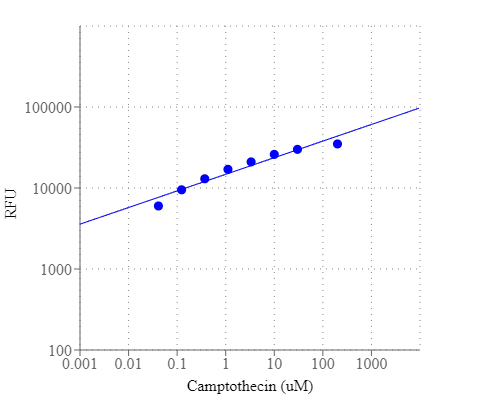
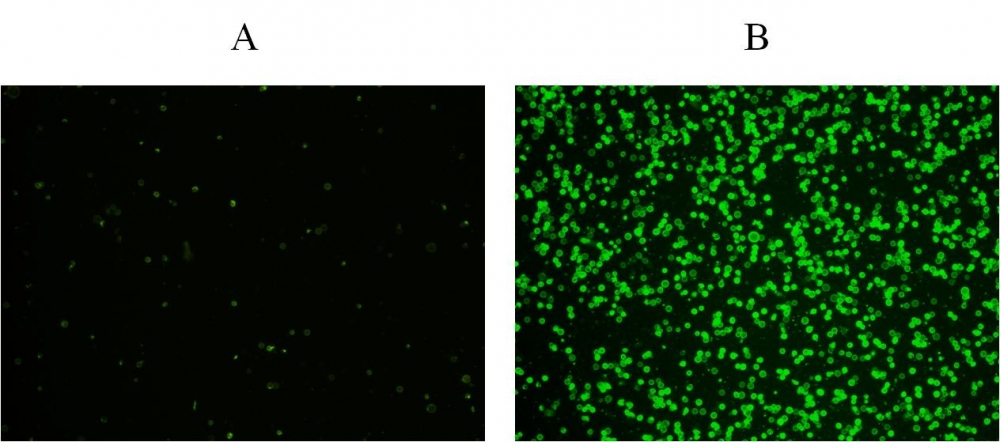
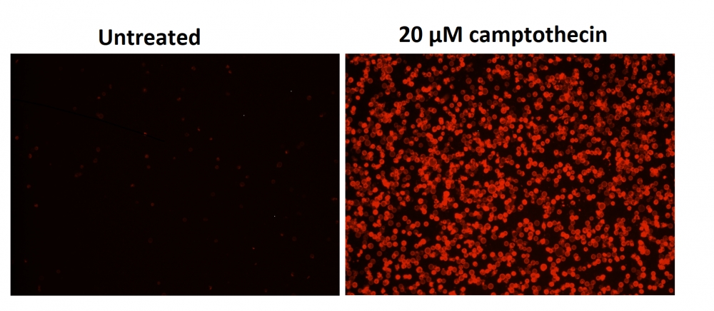
a.用2 µg / ml喜树碱处理Jurkat细胞3小时。
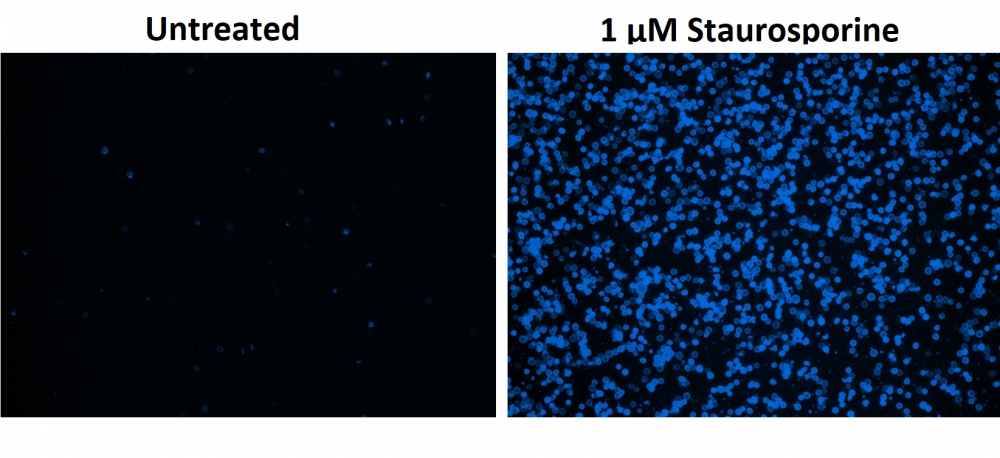
b.用1 µM星形孢菌素处理Jurkat细胞3小时。
c.用4 µg / ml喜树碱处理HL-60细胞4小时。
d.用1 µM星形孢菌素处理HL-60细胞4小时。 注意:应单独评估每种细胞系,以确定诱导凋亡的细胞密度。

2.以1:150的比例添加150X TF3-DEVD-FMK储备溶液和或以1:100的比例将Annexin V-iFluor 488 (组分B)添加到每个孔中。

3.将细胞在37°C,5%CO2培养箱中孵育1小时。 注意:对于TF3-DEVD-FMK标记,细胞可以浓缩至约5 X 10⁶细胞/ mL。 对于贴壁细胞,用0.5 mM EDTA轻轻提起细胞以保持细胞完整,并在与TF3-DEVD-FMK孵育之前用含血清的培养基洗涤一次。 合适的孵育时间取决于所用的单个细胞类型和细胞浓度。 优化每个实验的孵育时间。 贴壁细胞的膜联蛋白V流式细胞仪分析未经过常规测试,因为在细胞分离或收获过程中可能会发生特定的膜损伤。

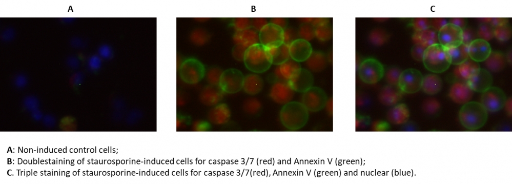
4.如果需要,用DNA染色剂标记细胞(例如,对于整个细胞核染色剂,使用Hoechst标记,或者如果仅用Annexin V-iFluor 488 标记细胞,则用碘化丙啶标记死细胞)。

5.以约200g的转速旋转细胞2分钟,并用1 mL(或使用96孔板的孔,每孔200 µL)洗涤细胞(组分E)洗涤两次。 用所需量的洗涤缓冲液重悬细胞。 注意:TF3-DEVD-FMK和Annexin V-iFluor 488 是荧光的,因此重要的是洗净任何未结合的试剂以消除背景。 对于分离的细胞,应将细胞浓度调整为每个酶标仪孔,2-5 X 10⁵细胞/ 100 µL等分试样。

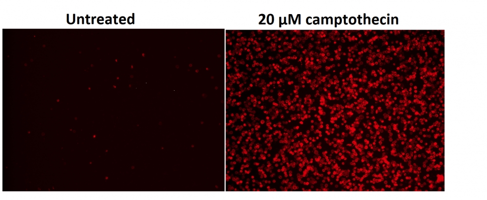
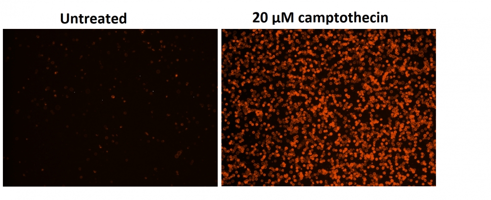
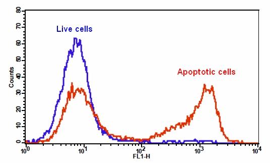
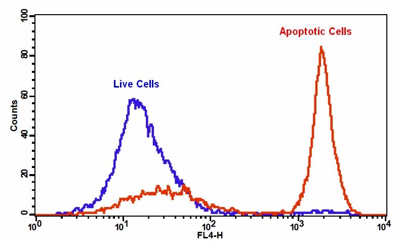
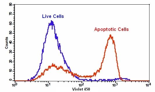
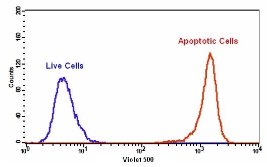
6.通过荧光显微镜,流式细胞仪或荧光酶标仪检测荧光强度,对于TF3-DEVD-FMK,Ex / Em = 550/595 nm;对于Annexin V-iFluor 488 ,490/525;对于Hoechst染色,350/461 nm;碘化丙锭为535/635。
对于流式细胞仪:使用膜联蛋白V-iFluor 488 的FL1通道,用于TF3-DEVD-FMK的FL2通道来检测荧光强度。

对于荧光显微镜:将100 µL细胞悬液放入黑色96孔板的每个孔中。 在荧光显微镜下观察细胞,使用TRITC通道用于TF3-DEVD-FMK,FITC通道用于Annexin V-iFluor 488 (TRITC通道用于碘化丙啶染色,DAPI通道用于Hoechst染色)。

对于荧光酶标仪:将100 µL细胞悬液放入黑色96孔板的每个孔中。 使用荧光酶标仪在Ex / Em = 490/525 nm(截止= 515 nm)或550/595 nm(截止= 570 nn)的Ex / Em = 490/525 nm(截止= 515 nm)处检测荧光强度(底部读取模式)适用于TF3-DEVD-FMK。注意:如果需要平衡细胞浓度,请调整诱导细胞的悬浮液体积,使其接近非诱导细胞的细胞密度。如果细胞治疗不会导致受刺激的细胞数量急剧减少,则此调整步骤是可选的。


参考文献

Anthocyanin-rich blackcurrant extract inhibits proliferation of the MCF10A healthy human breast epithelial cell line through induction of G0/G1 arrest and apoptosis
Authors: Nanashima, Naoki and Horie, Kayo and Chiba, Mitsuru and Nakano, Manabu and Maeda, Hayato and Nakamura, Toshiya
Journal: Molecular Medicine Reports (2017): 6134–6141

Clusterin signals via ApoER2/VLDLR and induces meiosis of male germ cells
Authors: Riaz, Muhammad Assad and Stammler, Angelika and Borgers, Mareike and Konrad, Lutz
Journal: American Journal of Translational Research (2017): 1266

Detecting Apoptosis, Autophagy, and Necrosis
Authors: Coleman, Jack and Liu, Rui and Wang, Kathy and Kumar, Arun
Journal: Apoptosis Methods in Toxicology (2016): 77–92

A pharmaceutical preparation of Salvia miltiorrhiza protects cardiac myocytes from tumor necrosis factor-induced apoptosis and reduces angiotensin II-stimulated collagen synthesis in fibroblasts
Authors: Ling S, Luo R, Dai A, Guo Z, Guo R, Komesaroff PA.
Journal: Phytomedicine (2009): 56

Agmatine protects cultured retinal ganglion cells from tumor necrosis factor-alpha-induced apoptosis
Authors: Hong S, Kim CY, Lee JE, Seong GJ.
Journal: Life Sci (2009): 28

Concurrent induction of necrosis, apoptosis, and autophagy in ischemic preconditioned human livers formerly treated by chemotherapy
Authors: Domart MC, Esposti DD, Sebagh M, Olaya N, Harper F, Pierron G, Franc B, Tanabe KK, Debuire B, Azoulay D, Brenner C, Lemoine A.
Journal: J Hepatol (2009): 881

Down-regulation of myeloid cell leukemia 1 by epigallocatechin-3-gallate sensitizes rheumatoid arthritis synovial fibroblasts to tumor necrosis factor alpha-induced apoptosis
Authors: Ahmed S, Silverman MD, Marotte H, Kwan K, Matuszczak N, Koch AE.
Journal: Arthritis Rheum (2009): 1282

Exaggerated up-regulation of tumor necrosis factor alpha-dependent apoptosis in the older mouse liver following reperfusion injury: targeting liver protective strategies to patient age
Authors: Selzner M, Selzner N, Chen L, Borozan I, Sun J, Xue-Zhong M, Zhang J, McGilvray ID.
Journal: Liver Transpl (2009): 1594

Increased apoptosis in HepG2.2.15 cells with hepatitis B virus expression by synergistic induction of interferon-gamma and tumour necrosis factor-alpha
Authors: Shi H, Guan SH.
Journal: Liver Int (2009): 349

Outside-to-inside signaling through transmembrane tumor necrosis factor reverses pathologic interleukin-1beta production and deficient apoptosis of rheumatoid arthritis monocytes
Authors: Meusch U, Rossol M, Baerwald C, Hauschildt S, Wagner U.
Journal: Arthritis Rheum (2009): 2612