线粒体膜电位荧光探针JC-1 CAS 3520-43-2-AAT Bioquest荧光染料

上海金畔生物科技有限公司代理AAT Bioquest荧光染料全线产品,欢迎访问AAT Bioquest荧光染料官网了解更多信息。
线粒体膜电位荧光探针JC-1 CAS 3520-43-2价格 1386
产品规格

5 mg

产品货号

线粒体膜电位荧光探针JC-1 CAS 3520-43-2

产品参数
Ex (nm) 515 Em (nm) 530
分子量 652.23 溶剂 DMSO
存储条件 在零下15度以下保存, 避免光照
产品概述

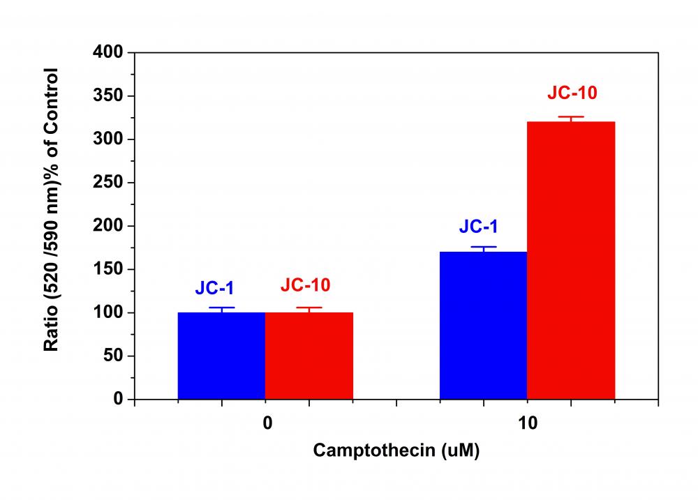
线粒体膜电位荧光探针JC-1是美国AAT Bioquest生产的用于线粒体膜电位检测的试剂,JC-1广泛用于通过流式细胞术确定线粒体膜电位。它能够选择性地进入线粒体,并且随着线粒体膜电位增加(超过约80-100mV的值)可逆地将其颜色从绿色变为橙色。这种性质是由于线粒体膜极化后JC-1聚集体的可逆形式导致发射光从530nm(即JC-1单体形式的发射)转变为590nm(即J-聚集形式的发射)。当在490nm处激发时,随着线粒体膜变得更加极化,JC-1的颜色从绿色橙色可逆地变为绿色橙色。使用通常安装在所有流式细胞仪中的滤波器可以检测两种颜色,可以在荧光通道1(FL1)中分析绿色发射,在通道2(FL2)中分析绿色橙色发射。使用JC-1的主要优点是,它可以从绿色到橙色荧光发射转移,它既可以定性,又可以在FL1和FL2通道中检测到纯荧光强度又可以定量。除了广泛用于流式细胞仪外,JC-1还用于荧光成像。我们已经开发出一种在荧光酶标仪平台中使用它的方案,尽管JC-1在许多实验室中被广泛使用,但其水溶性差使得它在某些应用中难以使用。我们的JC-10具有比JC-1更好的水溶性,并且JC-10在一些细胞系中具有优于JC-1的性能。金畔生物是AAT Bioquest 的中国代理商,为您提供优质的线粒体膜电位荧光探针。

点击查看光谱


适用仪器


流式细胞仪  
Ex: 488 nm
Em: 530/30 nm, 575/26 nm
通道: FITC, PE通道


荧光显微镜

Ex: 490 nm
Em: 525 nm(比率分析为 590 nm)
推荐孔板: 黑色透明底板
通道: FITC 和 TRITC通道


荧光酶标仪

 
Ex: 490 nm
Em: 525 nm(比率分析为 590 nm)
推荐孔板: 纯黑色孔板
实验方案

JC-1样品分析方案

概述

用测试化合物制备细胞

添加JC-1工作溶液(100μL/孔用于96孔板或25μL/孔用于384孔板)在室温或37℃孵育1小时

移除JC-1工作溶液

读取 Ex / Em = 490 / 525nm和490 / 590nm处的荧光强度

注意:以下是我们推荐的活细胞方案。该方案仅提供指南,实际情况应根据您的具体实验进行调整修改。


1.准备JC-1工作溶液:

1.1在高质量无水DMSO中制备2至10 mM JC-1的储备液。 应随用随配及时使用或等分配制,应将使用剩余的溶液等分并在<-20℃冷冻。

注意:避免反复冻融循环,避免光照。

1.2制备1X JC-1工作溶液:在实验当天,将JC-1固体溶解在DMSO中或将等分试样的JC-1储备溶液解冻至室温。在Hanks和20 mM Hepes缓冲液(HHBS)或其他缓冲液(pH 7)和0.02%Pluronic®F-127中制备10至30μM1XJC-1工作溶液。

注意:JC-1不溶于水,因此它会在溶液中聚集。 建议在将JC-1工作溶液加载到微孔板之前对其进行过滤。


2.用荧光酶标仪进行JC-1检测:

2.1用测试化合物细胞培养所需的一段时间(例如,Jurkat细胞可以用喜树碱处理4-6小时)以诱导细胞凋亡。对于空白孔(没有细胞的培养基),加入相应量的化合物缓冲液。

2.2将100μL/孔的96孔板或25μL/孔的384孔板的JC-1工作溶液(来自步骤1.2)加入到细胞板中。

2.3将JC-1加载在37℃,5%CO2培养箱中培养15-60分钟。

注意:适当的孵育时间取决于所使用的单个细胞类型和细胞浓度,每个实验的孵育时间需要根据相应实验进行控制。

2.4从平板上取下JC-1工作溶液,用HHBS或其他缓冲液清洗细胞。 将100μL/孔的96孔板或25μL/孔的384孔HHBS板加回到细胞板中。

2.5监测Ex / Em = 490 / 525nm和490 / 590nm处的荧光变化以进行比率分析。


3.用荧光显微镜或流式细胞仪进行JC-1测定:

3.1用测试化合物细胞培养所需的一段时间(例如,Jurkat细胞可以用喜树碱处理4-6小时)以诱导细胞凋亡。

3.2离心细胞,每管取1-5×10⁵个细胞。

3.3在500μLJC-1工作溶液中重悬细胞(来自步骤1.2)。

3.4在室温或37°C下避光孵育10至30分钟。

3.5用HHBS或其他缓冲液洗涤细胞。将细胞重悬于500μLHHBS中以获得每管1-5×10⁵个细胞。

3.6用荧光显微镜(使用FITC和TRITC滤光片)或流式细胞仪(使用FL1和FL2通道)观察Ex / Em = 490 / 525nm和490 / 590nm处的荧光变化。


参考文献

Cell death mechanisms of the anti-cancer drug etoposide on human cardiomyocytes isolated from pluripotent stem cells
Authors: Harshal Nemade, Umesh Chaudhari, Aviseka Acharya, Jürgen Hescheler, Jan Georg Hengstler, Symeon Papadopoulos, Agapios Sachinidis
Journal: Archives of Toxicology (2018): 1–18

Mesenchymal stem cells ameliorate hyperglycemia-induced endothelial injury through modulation of mitophagy
Authors: Wuzheng Zhu, Yujia Yuan, Guangneng Liao, Lan Li, Jingping Liu, Younan Chen, Jie Zhang, Jingqiu Cheng, Yanrong Lu
Journal: Cell Death & Disease (2018): 837

overexpression of protocadherin 7 inhibits neuronal survival by downregulating Birc5 in vitro
Authors: Huajuan Xiao, Ziling Sun, Jun Wan, Shengtao Hou, Yi Xiong
Journal: Experimental cell research (2018): 71–80

Therapeutic potential of GSK-J4, a histone demethylase KDM6B/JMJD3 inhibitor, for acute myeloid leukemia
Authors: Yunan Li, Mingying Zhang, Mengyao Sheng, Peng Zhang, Zizhen Chen, Wen Xing, Jie Bai, Tao Cheng, Feng-Chun Yang, Yuan Zhou
Journal: Journal of Cancer Research and Clinical Oncology (2018): 1–13

Hyaluronan polymeric micelles for topical drug delivery
Authors: Daniela Smejkalová, Tomáš Muthny, Kristina Nešporová, Martina Hermannová, Eva Achbergerová, Gloria Huerta-Angeles, Marek Svoboda, Martin Cepa, Veronika Machalová, Dominika Luptáková
Journal: Carbohydrate Polymers (2017): 86–96

New ruthenium compounds bearing semicarbazone 2-formylopyridine moiety: Playing with auxiliary ligands for tuning the mechanism of biological activity
Authors: Michal Lomzik, Olga Mazuryk, Dorota Rutkowska-Zbik, Grazyna Stochel, Philippe C Gros, Malgorzata Brindell
Journal: Journal of Inorganic Biochemistry (2017)

Inhibition of mTOR’s Catalytic Site by PKI-587 Is a Promising Therapeutic Option for Gastroenteropancreatic Neuroendocrine Tumor Disease
Authors: Helma Freitag, Friederike Christen, Florentine Lewens, Irina Grass, Franziska Briest, Sara Iwaszkiewicz, Britta Siegmund, Patricia Grabowski
Journal: Neuroendocrinology (2016)

Mesenchymal Stem Cells-Conditioned Media Ameliorates Diabetic Endothelial dysfunction by Improving Mitochondrial Bioenergetics via the Sirt1/AMPK/PGC-1α Pathway
Authors: Yujia Yuan, Meimei Shi, Lan Li, Jingping Liu, Bo Chen, Younan Chen, Xingxing An, Shuyun Liu, Ruixi Luo, Dan Long
Journal: Clinical Science (2016): CS20160235

mTOR complex-2 stimulates acetyl-CoA and de novo lipogenesis through ATP citrate lyase in HER2/PIK3CA-hyperactive breast cancer
Authors: Yaqing Chen, Jianchang Qian, Qun He, Hui Zhao, Lourdes Toral-Barza, Celine Shi, Xuesai Zhang, Jiang Wu, Ker Yu
Journal: Oncotarget (2016): 25224–25240

Multiple Active Compounds from Viscum album L. Synergistically Converge to Promote Apoptosis in Ewing Sarcoma
Authors: Monika Twardziok, Susann Kleinsimon, Jana Rolff, Sebastian Jäger, Angelika Eggert, Georg Seifert, Catharina I Delebinski
Journal: PloS one (2016): e0159749

线粒体膜电位荧光探针JC-10 JC-1的卓越代替品-AAT Bioquest荧光染料

上海金畔生物科技有限公司代理AAT Bioquest荧光染料全线产品,欢迎访问AAT Bioquest荧光染料官网了解更多信息。
线粒体膜电位荧光探针JC-10 JC-1的卓越代替品价格 1386
产品规格

5×100 uL

产品货号

线粒体膜电位荧光探针JC-10 JC-1的卓越代替品

产品参数
Ex (nm) 508 Em (nm) 524
分子量 583.34 溶剂 DMSO
存储条件 在零下15度以下保存, 避免光照
产品概述

线粒体膜电位荧光探针JC-10是美国AAT Bioquest研发的用于检测线粒体膜电位的荧光探针是JC-1的替代品。JC-1在许多实验室中被广泛使用,但其水溶性差使得它在某些应用中难以使用。即使在1μM浓度下,JC-1也倾向于在水性缓冲液中沉淀。当需要高染料浓度时,JC-10已被开发为JC-1的替代物。与JC-1相比,我们的JC-10具有更好的水溶性。 JC-10能够选择性地进入线粒体,并随着膜电位的增加可逆地将其颜色从绿色变为橙色。这种性质是由于膜极化时JC-10聚集体的可逆形成导致发射光从520nm(即JC-10单体形式的发射)转变为570nm(即J-聚集体形式的发射)。当在490nm激发时,随着线粒体膜变得更加极化,JC-10的颜色从绿色橙色可逆地变为绿色橙色。使用通常安装在所有流式细胞仪中的过滤器可以检测两种颜色。可以在荧光通道1(FL1)中分析绿色发射,在通道2(FL2)中分析绿色橙色发射。除了用于流式细胞仪外,JC-10还可用于荧光成像。我们已经开发出一种在荧光微孔板平台中使用JC-10的方案。在一些细胞系中,JC-10具有优于JC-1的性能。金畔生物是AAT Bioquest 的中国代理商,为您提供优质的线粒体膜电位荧光探针。

点击查看光谱

实验方案

JC-10的分析方案

概述

准备含有测试化合物的细胞

添加JC-10工作溶液(100μL/孔用于96孔板或25μL/孔用于384孔板)

在室温或37 ℃孵育1小时

在Ex读取荧光强度 / Em = 490 / 525nm和540 / 590nm

注意:以下是我们推荐的活细胞方案。 该协议仅提供指南,应根据您的特定需求进行修改。


操作步骤

1.准备JC-10工作溶液:

1.1每瓶DMSO原液(100μL,2 mg / mL,3 mM)只能使用一次。任何未使用的小瓶应储存在<-20℃。注意:避免反复冻融循环,并避光.

1.2准备1X JC-10工作溶液:在实验当天,解冻一份JC-10 stockolution到室温。在Hanks和20 mM Hepesbuffer(HHBS)或您选择的缓冲液(pH 7-8,含0.02%Pluronic F-127)中制备10至30μM1X工作溶液。通过votexing将它们混合均匀。注意:对于某些细胞系,pH值为8的工作溶液可能会阻止JC-10泄漏。

2.用荧光酶标仪进行JC-10检测:

2.1用测试化合物处理细胞一段所需的时间(例如,Jurkat细胞可以用喜树碱处理4-6小时)以诱导细胞凋亡。 对于空白孔(没有细胞的培养基),加入相应量的化合物缓冲液。

2.2将100μL/孔/ 96孔板或25μL/孔/ 384孔板的JC-10工作溶液(来自步骤1.2)加入到细胞板中。

2.3将JC-10加载板在37 oC,5%CO2培养箱中孵育15-60分钟。

注意:适当的孵育时间取决于所使用的单个细胞类型和细胞浓度。优化每个实验的孵育时间。

2.4监测Ex / Em = 490/525 nm(FITC通道)和540/590 nm(TRITC通道)的荧光变化,进行比率分析。

可选:从板上取下JC-10工作溶液; 在分析之前,将100μL/孔/ 96孔板或25μL/孔/ 384孔HHBS板加回到细胞板中。

3.用荧光显微镜或流式细胞仪进行JC-10检测:

3.1用测试化合物处理细胞一段所需的时间(例如,Jurkat细胞可以用喜树碱处理4-6小时)以诱导细胞凋亡。

3.2离心细胞,每管取1-5×10⁵个细胞。

3.3将细胞重悬于500μLJC-10工作溶液中(来自步骤1.2)。

3.4在室温或37°C,5%CO2培养箱中孵育10至30分钟,避光。

注意:适当的孵育时间取决于所使用的单个细胞类型和细胞浓度。优化每个实验的孵育时间。

3.5使用荧光显微镜(使用FITC和TRITC过滤器)或流式细胞仪(使用FL1和FL2通道)监测Ex / Em = 490/525 nm和540/590 nm处的荧光变化。可选:移除JC-10工作 从板上解决; 在荧光显微镜下分析之前,将100μL/孔/ 96孔板或25μL/孔/ 384孔HHBS板加回到细胞板中。


参考文献

JC-10™ has been used to study many biologically significant processes across several key disciplines. To list a few, JC-10™ has been used to investigate topics such as mitochondrial membrane potential, cytotoxicity, cell viability, oxidative stress, cancer metastasis, apoptosis, signal transduction, mitochondrial fission and induced pluripotent stem cells (iPSCs). 

Below, you may find a small sampling of specific JC-10™ applications. To inquire about a potential application of JC-10™, or to consult with our fluorescent dye specialists, please contact us at support@aatbio.com or 1-800-990-8053.

High-content assays for hepatotoxicity using induced pluripotent stem cell–derived cells.
Researchers use JC-10™ to monitor mitochondrial depolarization as an indication of hepatotoxicity and oxidative stress in induced pluripotent stem cell-derived cells, with the ultimate goal of designing a reliable, high-content and imaging-based in vitro toxicity assay. 

High-Content High-Throughput Assays for Characterizing the Viability and Morphology of Human iPSC-Derived Neuronal Cultures
JC-10™ was used to study neurons derived from induced pluripotent stem-cells. Since JC-10™ will accumulate in the mitochondria of viable cells, it was used to determine cell viability in a high-throughput assay context.

Anticancer Activity of New Synthetic α-Methylene-δ-Lactones on Two Breast Cancer Cell Lines
Researchers chose JC-10™ to investigate mitochondrial membrane potential and membrane integrity in cells treated with natural products, such as α-Methylene-δ-Lactones, with the goal of developing new treatments for breast cancer.

Tetrandrine protects mouse retinal ganglion cells from ischemic injury.
JC-10™ was used in flow cytometry to study mitochondrial membrane potential (ΔΨm) in primary cultured retinal ganglion cells, as an extension of the field of drug discovery into prevention of ischemic injury. 

Midazolam induces cellular apoptosis in human cancer cells and inhibits tumor growth in xenograft mice
In a study of human cancer cells, JC-10™ was employed to track cellular apoptosis as a function of mitochondrial membrane potential, and consequently, mitochondrial activity. Researchers were interested in the possible anesthetic properties of midazolam for anticancer drug delivery.

Mitochondrial proteomics with siRNA knockdown to reveal ACAT1 and MDH2 in the development of doxorubicin-resistant uterine cancer
JC-10™ was used by researchers for the purposes of drug discovery. In particular, researchers were interested in finding new treatments for doxorubicin-resistant uterine cancer, using JC-10™ to monitor mitochondrial membrane potential and validate cell viability results. 

Cold exposure lowers energy expenditure at the cellular level
Researchers used JC-10™ to investigate the relationship between temperature and cellular activity. In particular, researchers wanted to explore if cold temperature acts as a stressor on mitochondrial membrane potential, with regards to the oxidative phosphorylation process which generates ATP.

Susceptibility of gametes and embryos of the eastern oyster, Crassostrea virginica, to Karenia brevis and its toxins
JC-10™ was used in the study of sperm viability, fertilization successs and embryonic survival of Crassostrea virginica. Specifically, JC-10™ was used to quantify mitochondrial membrane potential in sperm cells and to determine possible toxicity effects of algal blooms.

Calmodulin antagonists induce cell cycle arrest and apoptosis in vitro and inhibit tumor growth in vivo in human multiple myeloma
JC-10™ was used by researchers to study cell cycle and apoptosis in human multiple myeloma. JC-10™ functioned as a probe for the detection of mitochondrial membrane potential depolarization, which was crucial to the study of caspase activated apoptosis.

Activation of the mitochondrial apoptotic pathway produces reactive oxygen species and oxidative damage in hepatocytes that contribute to liver tumorigenesis
Researchers were interested in the pathways involved with liver tumorigenesis. To that end, they used JC-10™ to study activation of apoptotic pathways related to changes in mitochondrial membrane potential, with the ultimate goal of discovering if antioxidant therapy might help suppress liver carinogenesis.