
上海金畔生物科技有限公司代理AAT Bioquest荧光染料全线产品,欢迎访问AAT Bioquest荧光染料官网了解更多信息。
Cy5DIGE NHS ester价格 7092
产品规格
100 nmoles
产品货号
产品参数
| Ex (nm) | 651 | Em (nm) | 670 |
| 分子量 | 707.65 | 溶剂 | DMSO |
| 存储条件 | 在零下15度以下保存, 避免光照 |
产品概述
产品基本信息
产品名称:Cy5DIGE NHS ester
储存条件:-15℃避光防潮
保质期:24个月
产品物理化学光谱特性
Ex:650nm
Em:669nm
溶剂:DMSO
产品介绍
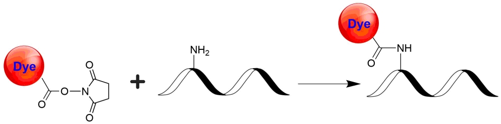
Cy5DIGE NHS酯等同于Cy5®NHS酯染料,是用于标记DIGE蛋白分析的主要染料之一。Cy5DIGE通常与Cy2DIGE和Cy3DIGE一起使用。Cy2DIGE,C3DIGE和Cy5DIGE是专为比较两个或三个裂解液样品中蛋白质表达而设计的。匹配迁移率和明亮的染料荧光使2D凝胶电泳能够有效检测和微量蛋白质的高分辨率分离。Cy2DIGE,C3DIGE和Cy5DIGE与所有能够检测Cy2,Cy3和Cy5的染料成像仪兼容。Cy2DIGE,C3DIGE和Cy5DIGE标记的凝胶是规格匹配、电荷匹配的荧光染料,用于检测二维荧光差异凝胶电泳(DIGE)中的蛋白质丰度差异。Cy2DIGE C3DIGE和Cy5DIGE在同一2-D电泳凝胶上多可检测三个预先标记的蛋白质样品和标准品。规格和电荷匹配的染料可以使标记的样品在凝胶内共迁移。它们在标记的蛋白质上很亮,可以在标记、分离和扫描过程中将信号损失降至低。这些染料的光谱重叠很少,可将通常导致高背景的串扰降至低。它们对pH值不敏感的荧光使DIGE可以在较宽的pH范围内运行。金畔生物是AAT Bioquest的中国代理商,为您提供优质的Cy5DIGE NHS ester。
点击查看光谱
参考文献
LncRNA AL592284. 1 facilitates proliferation and metastasis of cervical cancer cells via miR-30a-5p/Vimentin/EMT axis
Authors: Zhang, Jing and Liu, Hong-li and Liu, Jing-bo and Zhang, Yuan and Liu, Jing and Li, Yan-hua
Journal: Biochemical and Biophysical Research Communications (2021): 95–102
Authors: Zhang, Jing and Liu, Hong-li and Liu, Jing-bo and Zhang, Yuan and Liu, Jing and Li, Yan-hua
Journal: Biochemical and Biophysical Research Communications (2021): 95–102
Early quantitative profiling of differential retinal protein expression in lens-induced myopia in guinea pig using fluorescence difference two-dimensional gel electrophoresis
Authors: Wu, Y., Lam, C. S., Tse, D. Y., To, C. H., Liu, Q., McFadden, S. A., Chun, R. K., Li, K. K., Bian, J., Lam, C.
Journal: Mol Med Rep (2018): 5571-5580
Authors: Wu, Y., Lam, C. S., Tse, D. Y., To, C. H., Liu, Q., McFadden, S. A., Chun, R. K., Li, K. K., Bian, J., Lam, C.
Journal: Mol Med Rep (2018): 5571-5580
Proteomic Analysis of Bovine Pregnancy-specific Serum Proteins by 2D Fluorescence Difference Gel Electrophoresis
Authors: Lee, J. E., Lee, J. Y., Kim, H. R., Shin, H. Y., Lin, T., Jin, D. I.
Journal: Asian-Australas J Anim Sci (2015): 788-95
Authors: Lee, J. E., Lee, J. Y., Kim, H. R., Shin, H. Y., Lin, T., Jin, D. I.
Journal: Asian-Australas J Anim Sci (2015): 788-95
Comparative proteomic analysis of different Toxoplasma gondii genotypes by two-dimensional fluorescence difference gel electrophoresis combined with mass spectrometry
Authors: Zhou, D. H., Zhao, F. R., Nisbet, A. J., Xu, M. J., Song, H. Q., Lin, R. Q., Huang, S. Y., Zhu, X. Q.
Journal: Electrophoresis (2014): 533-45
Authors: Zhou, D. H., Zhao, F. R., Nisbet, A. J., Xu, M. J., Song, H. Q., Lin, R. Q., Huang, S. Y., Zhu, X. Q.
Journal: Electrophoresis (2014): 533-45
Comparative proteomic analysis of Dan’er malts produced from distinct malting processes by two-dimensional fluorescence difference in gel electrophoresis (2D-DIGE)
Authors: Li, X., Jin, Z., Gao, F., Lu, J., Cai, G., Dong, J., Yu, J., Yang, M.
Journal: J Agric Food Chem (2014): 9310-6
Authors: Li, X., Jin, Z., Gao, F., Lu, J., Cai, G., Dong, J., Yu, J., Yang, M.
Journal: J Agric Food Chem (2014): 9310-6
Proteomic analysis of the hippocampus in Alzheimer’s disease model mice by using two-dimensional fluorescence difference in gel electrophoresis
Authors: Takano, M., Yamashita, T., Nagano, K., Otani, M., Maekura, K., Kamada, H., Tsunoda, S., Tsutsumi, Y., Tomiyama, T., Mori, H., Matsuura, K., Matsuyama, S.
Journal: Neurosci Lett (2013): 85-9
Authors: Takano, M., Yamashita, T., Nagano, K., Otani, M., Maekura, K., Kamada, H., Tsunoda, S., Tsutsumi, Y., Tomiyama, T., Mori, H., Matsuura, K., Matsuyama, S.
Journal: Neurosci Lett (2013): 85-9
Application of fluorescence two-dimensional difference in-gel electrophoresis as a proteomic biomarker discovery tool in muscular dystrophy research
Authors: Carberry, S., Zweyer, M., Sw and ulla, D., Ohlendieck, K.
Journal: Biology (Basel) (2013): 1438-64
Authors: Carberry, S., Zweyer, M., Sw and ulla, D., Ohlendieck, K.
Journal: Biology (Basel) (2013): 1438-64
Two-dimensional fluorescence difference gel electrophoresis analysis of Listeria monocytogenes submitted to a redox shock
Authors: Ignatova, M., Guevel, B., Com, E., Haddad, N., Rossero, A., Bogard, P., Prevost, H., Guillou, S.
Journal: J Proteomics (2013): 13-27
Authors: Ignatova, M., Guevel, B., Com, E., Haddad, N., Rossero, A., Bogard, P., Prevost, H., Guillou, S.
Journal: J Proteomics (2013): 13-27
Identification of new pathogenic candidates for diabetic macular edema using fluorescence-based difference gel electrophoresis analysis
Authors: Hern, undefined and ez, C., Garcia-Ramirez, M., Colome, N., Corraliza, L., Garcia-Pascual, L., Casado, J., Canals, F., Simo, R.
Journal: Diabetes Metab Res Rev (2013): 499-506
Authors: Hern, undefined and ez, C., Garcia-Ramirez, M., Colome, N., Corraliza, L., Garcia-Pascual, L., Casado, J., Canals, F., Simo, R.
Journal: Diabetes Metab Res Rev (2013): 499-506
Discovery and target identification of an antiproliferative agent in live cells using fluorescence difference in two-dimensional gel electrophoresis
Authors: Park, J., Oh, S., Park, S. B.
Journal: Angew Chem Int Ed Engl (2012): 5447-51
Authors: Park, J., Oh, S., Park, S. B.
Journal: Angew Chem Int Ed Engl (2012): 5447-51